


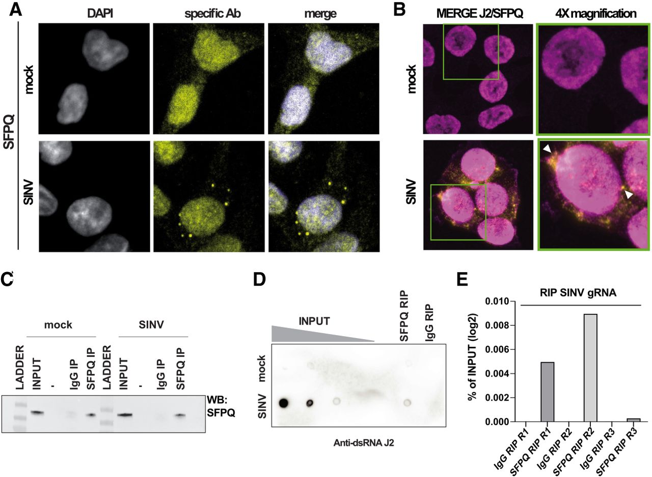
SFPQ binds to dsRNA and viral dsRNA upon SINV infection. (A) 4× magnification of the confocal immunofluorescence images shown in Supplemental Figure S4A. Anti-SFPQ mouse antibody, yellow signal. DAPI was used to stain the nuclei (in blue in the merge). (B) Confocal coimmunofluorescence analysis on mock and SINV WT infected HCT116 cells. Merge of anti-SFPQ rabbit antibody (in purple) or anti-dsRNA J2 mouse antibody (in yellow) is shown. Single antibodies and DAPI staining are shown in Supplemental Figure S4B. Magnification 63× (3×). Green squares: 4× higher magnification White arrows indicate J2 and SFPQ colocalization. (C) SFPQ immunoprecipitation on mock and SINV-infected HCT116 samples analyzed by western blot with anti-SFPQ rabbit antibody. (D) Dot blot revealed using the anti-dsRNA J2 antibody on RNA extracted from total lysate (INPUT, four 10-fold dilutions), SFPQ- or IgG control-RIP on mock (upper part) and SINV-infected (lower part) HCT116 cells. (E) RT-qPCR on SFPQ-IP and IgG CTRL-IP samples by using oligos to amplify SINV genomic RNA. The RNA enrichment upon SFPQ-IP or IgG IP is shown for three biological replicates and is expressed as percentage (%) of the INPUT (total RNA). The IgG RIP represent the background signal.




