


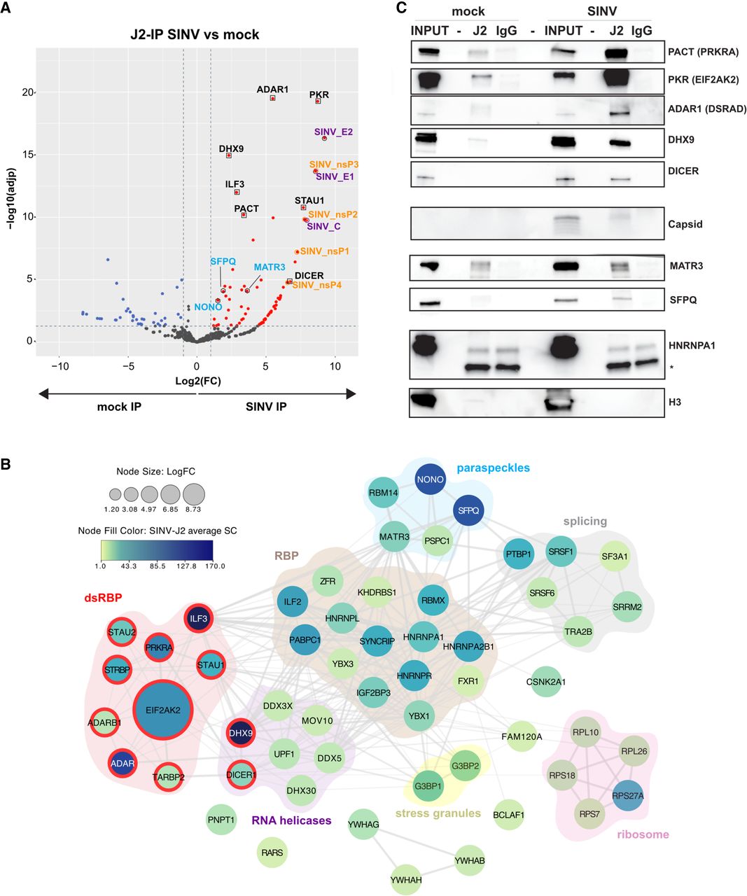
The network of dsRNA enriched proteins upon SINV infection in HCT116 cells. (A) Volcano plot showing the global enrichment of proteins upon dsRNA-IP (J2-IP) in SINV-infected versus mock conditions using DRIMS data from three replicates. Infection conditions: 24 h at MOI of 0.01. Red and blue dots represent proteins which are significantly enriched or depleted [adjusted P-value <0.05, a minimum of five SpC in the most abundant condition, and abs(Log2FC) > 1], respectively, in the infected samples compared to the uninfected ones. Viral nonstructural and structural proteins are indicated in orange and purple, respectively. Known dsRNA binding proteins are indicated in black and by a black square. Proteins present in paraspeckles are indicated in light blue and by a black hexagon. (B) Visualization of the interaction network of proteins identified by DRIMS upon infection, generated with the Cytoscape StringApp. The confidence score of each interaction is mapped to the edge thickness and opacity. The size of the node relates to the enrichment in log2 fold change (LogFC) over the J2-IP mock control. The protein abundance in the SINV-J2 IP sample is illustrated by a color scale and corresponds to the specific spectral count. Proteins were clustered into different functional categories using STRING enriched terms as a guideline. Proteins with dsRNA binding domain according to STRING enrichment (category: SMART domains) are highlighted in red. (C) Western blots performed on total lysate (INPUT), J2- or IgG-IP in mock and SINV-GFP-infected cells using the indicated specific antibodies. Histone 3 (H3) and HNRNPA1 were used as negative controls. (*) corresponds to an unspecific band.




