


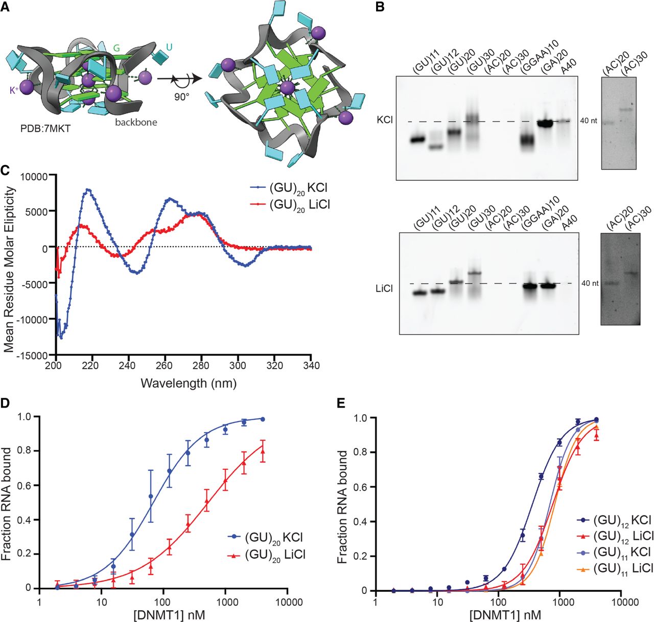
DNMT1 binds with high affinity to pUG-fold RNA. (A) Schematic of a pUG-fold. G and U residues are labeled in green and cyan, respectively. The phosphate backbone is highlighted in gray, and coordinated K+ ions are indicated by purple spheres. PDB: 7MKT. (B) Native gel of indicated RNAs with either 100 mM KCl or LiCl included in the folding buffer, the gel running buffer, and the gel itself. (A)40 was used as a marker for an unstructured single-stranded RNA (dashed line). Longer exposure of (AC)20 and (AC)30 staining is shown to the right, due to the low staining of AC sequences by SybrGold. (C) Circular dichroism spectra of (GU)20 in 100 mM KCl (blue) and LiCl (red). (D) Binding curves of (GU)20 bound to DNMT1 in 100 mM KCl (blue) and LiCl (red). (E) Binding curves of (GU)12 and (GU)11 bound to DNMT1 in 100 mM KCl (blue and light purple, respectively) and LiCl (red and orange, respectively). In D and E, points represent mean values and error bars represent SD for n = 3 replicates.




