


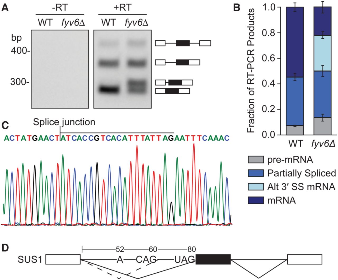
Loss of Fyv6 results in the use of an alternative 3′ SS in SUS1. (A) Representative gel image of RT-PCR of SUS1 in strains with (WT) and without (fyv6Δ) Fyv6 in a upf1Δ background. (+RT reactions contain reverse transcriptase; −RT control reactions do not contain reverse transcriptase). (B) Quantification of band intensities of each isoform as a fraction of the total SUS1 product in a lane. The bars indicate the average of three experiments with standard deviation. (C) Portion of the Sanger sequencing chromatogram of the SUS1 splice variant identified as an RT-PCR product in the fyv6Δ strain. The bar above the nucleotides indicates those from intron 1 included in the splice variant due to the use of an alternative 3′ SS. (D) Diagram of the SUS1 gene structure with the BS adenosine and the two, alternative 3′ SS of intron 1 indicated. The numbering of the nucleotides begins at the first nucleotide of intron 1. The newly identified 3′ SS is at position 60.




