


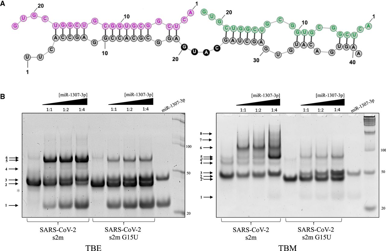
(A) Predicted miR-1307-3p binding sites to the s2m. The two miR-1307-3p molecules are shown in purple and green, respectively, whereas s2m is shown in gray. (B) Nondenaturing TBE (left) and TBM (right) gels of s2m and s2m G15U binding to miR-1307-3p in 1:1 and 1:2 and 1:4 s2m:miR ratios in the presence of 1 mM MgCl2. In TBE, both s2m elements prefer their monomeric states (arrow 2), whereas the miR-1307-3p exists as a monomer and dimer (arrows 1 and 3). As miR is added, upper complexes are formed between the two (arrows 4 and 5). Arrow 6 reveals the presence of the ED of the s2ms. Overall, the band intensity of the complexes formed by s2m G15U with miR-1307-3p is reduced. In TBM conditions, the s2m reference can form a stable complex with two miR-1307-3p (arrow 5) and a complex formed by two s2m each bound to one miR-1307-3p is also apparent (arrow 6). Arrows 7 and 8 indicate higher molecular weight complex that could be potentially formed by RNA LEGO type interactions mediated by the palindromic sequence GUGC at the 3′ end of miR-1307-3p interacting with another miR, as it is bound to a s2m. Arrows 4 and 4′ show the KD and duplex formed by the s2m reference, respectively. The monomers of s2m and s2m G15U migrate differently (arrows 2 and 2′), as described in the text.




