


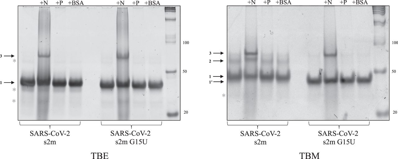
Nondenaturing gel electrophoresis of the s2m reference and s2m G15U converted to an ED structure by the viral N protein. Both s2m elements were incubated with 1 µM MgCl2 for 30 min, an additional 30 min with the N protein, and 5 min with proteinase K to digest the proteins prior to the samples being split in TBE (left) and TBM (right) conditions for electrophoresis. In both cases, the s2m remains preferentially monomeric (arrows 1 and 1′). The N protein converted both elements to an ED structure (arrow 3), and in the TBM gel, the N protein also stabilized the KD of the s2m reference (right, arrow 2). However, the band corresponding to the KD of the s2m G15U sample is still not observed. The P protein and BSA were used as controls and had no effect on the conversion of either s2m KD to the duplex conformation.




