


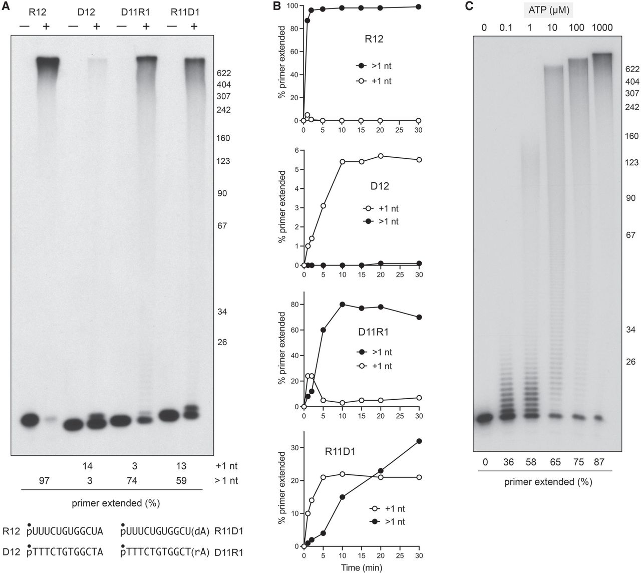
Primer specificity for RNA versus DNA. (A) Reaction mixtures (10 µL) containing 50 mM Tris-HCl (pH 7.4), 50 mM NaCl, 1 mM MnCl2, 0.5 mM DTT, 1 mM ATP, 0.1 µM (1 pmol) of the indicated 5′-32P labeled primer (R12—RNA; D12—DNA; D11R1—DNA with terminal 3′ ribonucleotide; R11D1—RNA with terminal 3′ deoxynucleotide), and either no added enzyme (−) or 0.1 µM (1 pmol) Pla1 were incubated at 30°C for 30 min and subsequently quenched. The sequence of the primers is shown at the bottom where p marked with (•) indicates the position of 32P-label. Primer extension products were resolved on 8% urea-PAGE and visualized by autoradiography. The size in nucleotides of a DNA ladder is indicated on the right. The radioactivity was quantified in ImageQuant-TL after scanning the gel with a Typhoon FLA7000 imager. (B) Reaction mixtures (85 µL) containing 50 mM Tris-HCl (pH 7.4), 50 mM NaCl, 1 mM MnCl2, 0.5 mM DTT, 1 mM ATP, 0.1 µM of the indicated 5′-32P labeled primer, and 0.04 µM Pla1 were incubated at 30°C. At times specified, 10 µL aliquots (containing 400 fmol Pla1, and 1 pmol primer and/or primer extension products) were withdrawn and quenched immediately by adjustment to 47.5% formamide/12.5 mM EDTA. The proportion of the extended primer, either by 1 nt or >1 nt, was quantified in ImageQuant-TL after scanning the gel with a Typhoon FLA7000 imager and is plotted as a function of time (min) for the indicated primer. (C) Reaction mixtures (10 µL) containing 50 mM Tris-HCl (pH 7.4), 50 mM NaCl, 1 mM MnCl2, 0.5 mM DTT, 0.1 µM 5′-32P labeled 12-mer RNA primer, 0.04 µM Pla1, and 0, 0.1, 1, 10, 100, or 1000 µM ATP as specified were incubated at 30°C for 30 min. The products were resolved by urea-PAGE, visualized by autoradiography, and the extents of primer extension were quantified by scanning the gel.




