


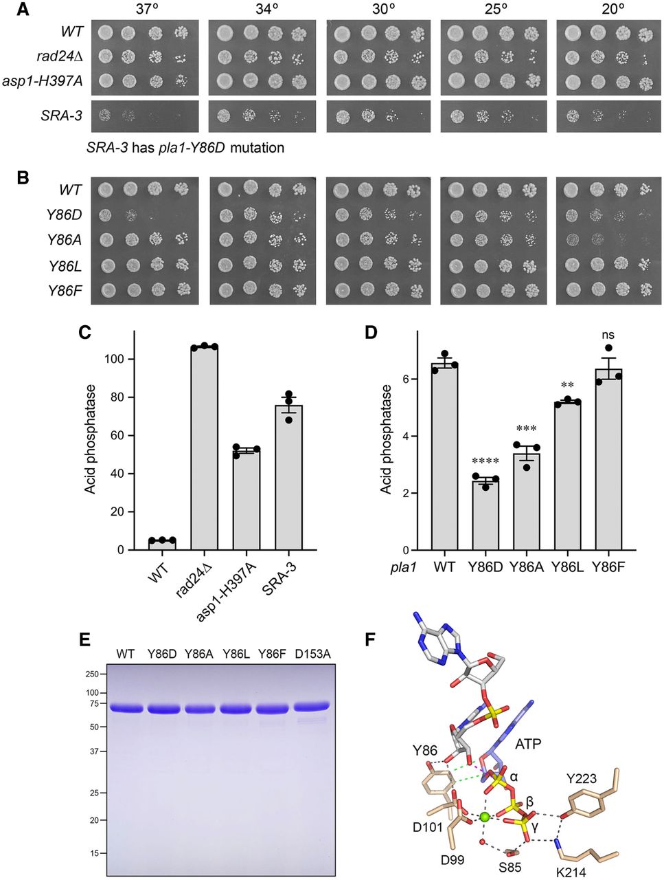
SRA screen identifies a novel mutation in poly(A) polymerase Pla1. (A,B) Serial dilutions of the indicated fission yeast strains were spotted on YES agar and grown at the temperatures specified. (C,D) The indicted strains were grown to A600 of 0.5–0.8 in liquid culture in YES medium at 30°C. Cells were then harvested, washed with water, and assayed for Pho1 acid phosphatase activity by conversion of p-nitrophenylphosphate to p-nitrophenol. Activity is expressed as the ratio of A410 (p-nitrophenol production) to A600 (input cells). (E) Aliquots (5 µg) of purified recombinant wild-type Pla1 and the indicated mutants were analyzed by SDS–PAGE. The Coomassie-blue stained gel is shown. The positions and sizes (kDa) of marker polypeptides are indicated on the left. (F) View of the active site of budding yeast poly(A) polymerase (D154A mutant) in complex with ATP (stick model with blue carbon and yellow phosphorus atoms), magnesium (green sphere), and ApAOH primer (stick model with gray carbons) (pdb 2Q66). Selected amino acids are shown as stick models with beige carbons and numbered according to their conserved counterparts in fission yeast Pla1. Hydrogen bonds and contacts to magnesium are indicated by black dashed lines. Van der Waals contacts of Tyr86 to the ATP ribose are denoted by green dashed lines. The trajectory of the nucleophilic attack of the ApA primer O3′ on the ATP α-phosphorus is indicated by a magenta dashed line.




