


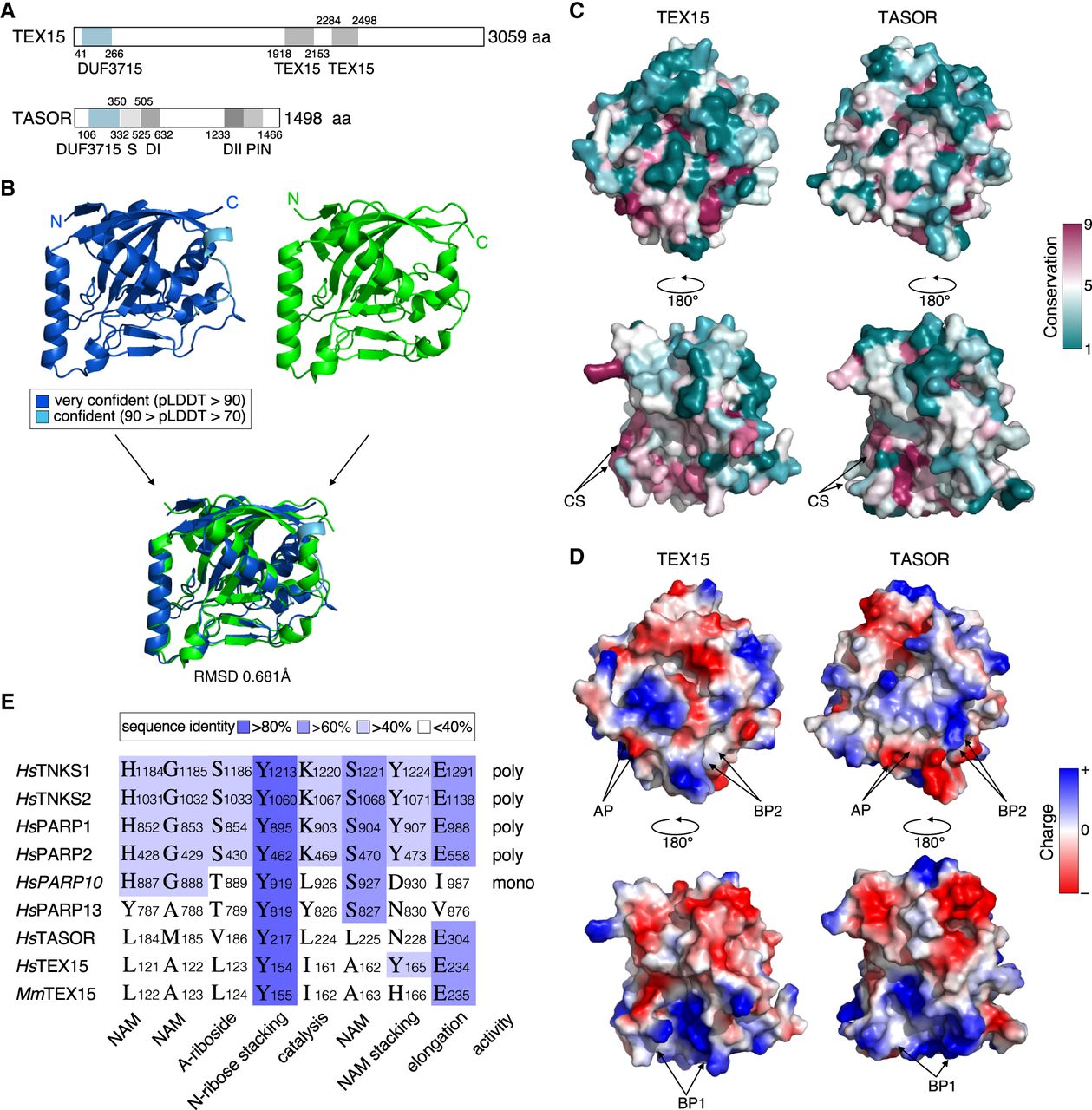
The DUF3715 domain of TASOR and TEX15 share extensive structural homology. (A) Schematic representation of TEX15 and TASOR domain structures. Characterized domains are in gray. (S) SPOC domain, (DI) DomI domain, (DII) DomII domain. (B) Structural prediction of TEX15 DUF3715 (colored by confidence [pLDDT] as indicated), experimental structure of TASOR DUF3715 (green) (PDB ID 6TL1 [Douse et al. 2020]) separately and aligned. (C) Surface views of TEX15 and TASOR as indicated from two different viewpoints via a 180° rotation. Color indicates the degree of conservation (magenta = conserved; teal = variable) across species. (CS) conserved patch. (D) Color indicates surface charge. ±64 ekT and ±66 ekT for TEX15 and TASOR, respectively. (AP) Acidic patch, (BP) basic patch. (E) Alignment of active PARP sites and NAD+ binding residues between PARP domain of selection of PARP family members and DUF3715 of TEX15 and TASOR. (NAM) Nicotinamide, (A-riboside) adenosyl-riboside, (N-riboside) nicotinamide-riboside. Sequence identity indicated in blue.




