


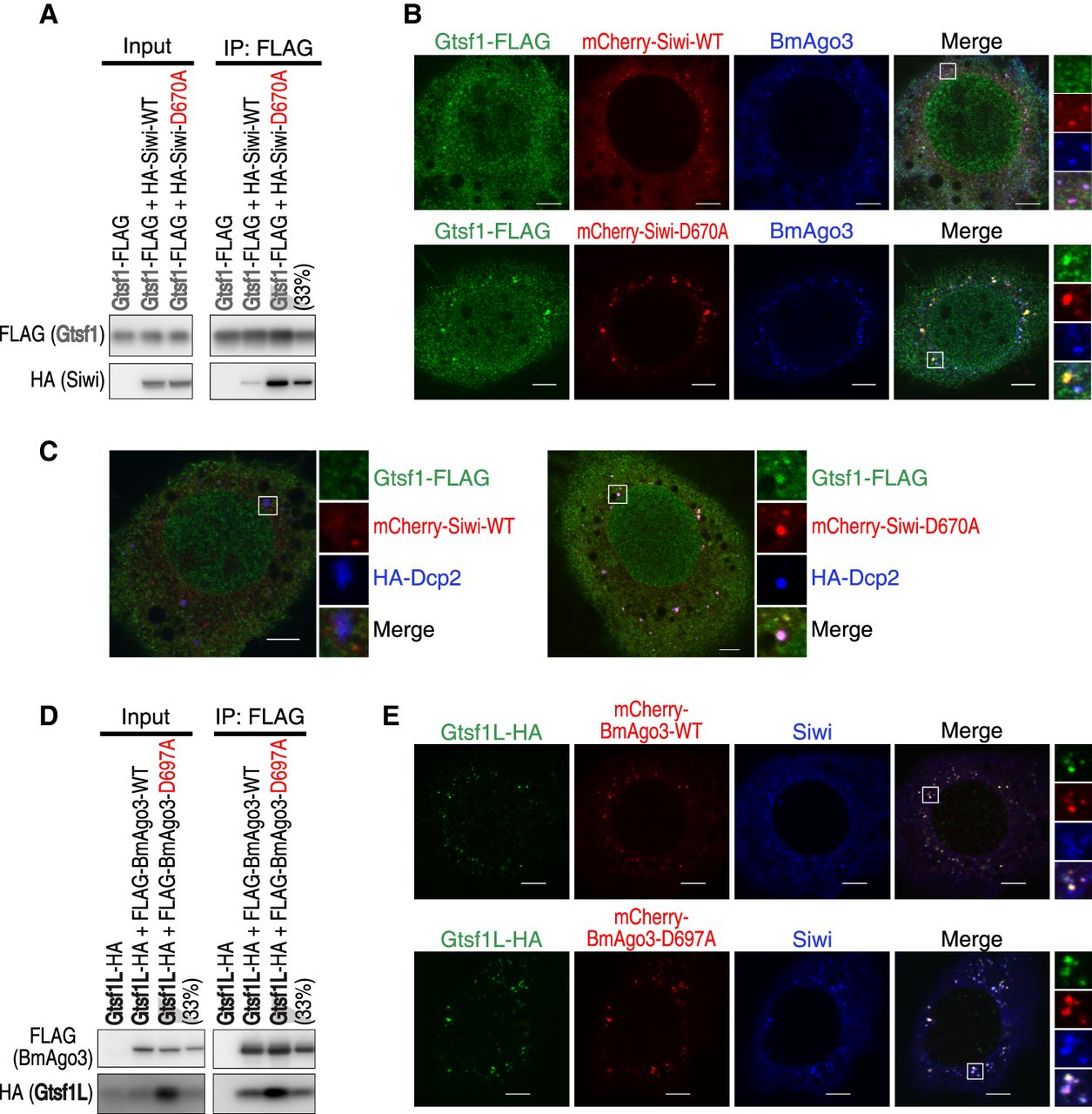
Gtsf1 and Gtsf1L show increased association with catalytically inactive Siwi and BmAgo3, respectively. (A) Western blot analysis of the interaction between Gtsf1-FLAG and HA-tagged wild-type (WT) Siwi or catalytically inactive Siwi-D670A. (B) Subcellular localization of Gtsf1-FLAG coexpressed with mCherry-Siwi or -Siwi-D670A. Scale bar, 5 µm. (C) Subcellular localization of Gtsf1-FLAG coexpressed with mCherry-Siwi or -Siwi-D670A, and HA-Dcp2, a P-body marker. Scale bar, 5 µm. (D) Western blot analysis of the interaction between Gtsf1L-HA and FLAG-tagged wild-type (WT) BmAgo3 or catalytically inactive BmAgo3-D697A. (E) Subcellular localization of Gtsf1L-HA coexpressed with mCherry-BmAgo3 or -BmAgo3-D697A. Scale bar, 5 µm.




