


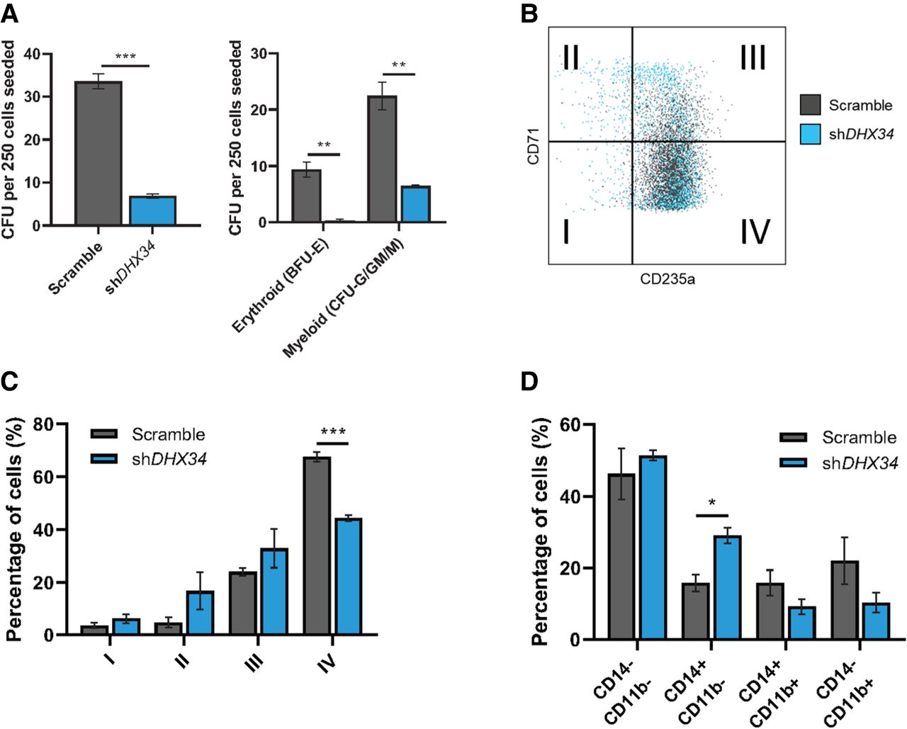
Loss of DHX34 impairs HSPC differentiation. (A) Cells were sorted by FACS (Dapi−CD34+GFP+) and placed in methylcellulose prior to being scored at day 14. Left panel represents total colonies scored, and right panel colonies scored in erythroid (BFU-E) and myeloid (CFU-G/GM/M) lineages (n = 3). (B) FACS plot representing cells at day 14 in erythroid conditions, blue represents cells KD for DHX34, and gray represents control cells (scramble). (C) Bar chart quantifying the percentage of erythroid differentiated cells in the different quadrants (I, II, III, and IV displayed on panel B). Cells were sorted by flow cytometer (Dapi−CD34+GFP+), cultured in erythroid differentiation conditions and immunophenotyped by FACS at day 14 based on CD71 and CD235a expression (n = 3). (D) Bar charts representing the percentage of granulo–monocytic cells after Dapi–CD34+GFP+ cells were sorted by flow cytometer and cultured in granulo–monocytic conditions for 2 wk. At day 14, cells were immunophenotyped by FACS based on CD11b and CD14 expression (n = 3). (*) P < 0.05; (**) P < 0.01; (***) P < 0.005.




