


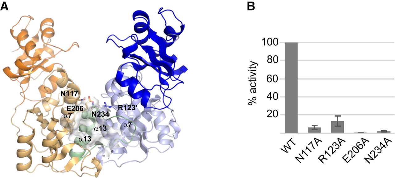
3D structure of YsgA. (A) Overall structure of the wild-type YsgA dimer, represented as cartoon. The subunits A and B are colored in blue and orange, respectively, with the amino-terminal domain in a darker color and residues 222–248 of the B subunit in green. The secondary structure elements mentioned in the manuscript are indicated. The cofactor-binding pocket in the B subunit is shown as a van der Waals surface. The single quote symbol refers to the dimerization partner. The side chain of the residues which were mutated to alanine (see panel B) are represented as sticks. (B) Residual methyltransferase activity of several YsgA mutants. The values are the percentage of activity compared to the wild-type enzyme and represent the mean ± SEM of four experiments.




