


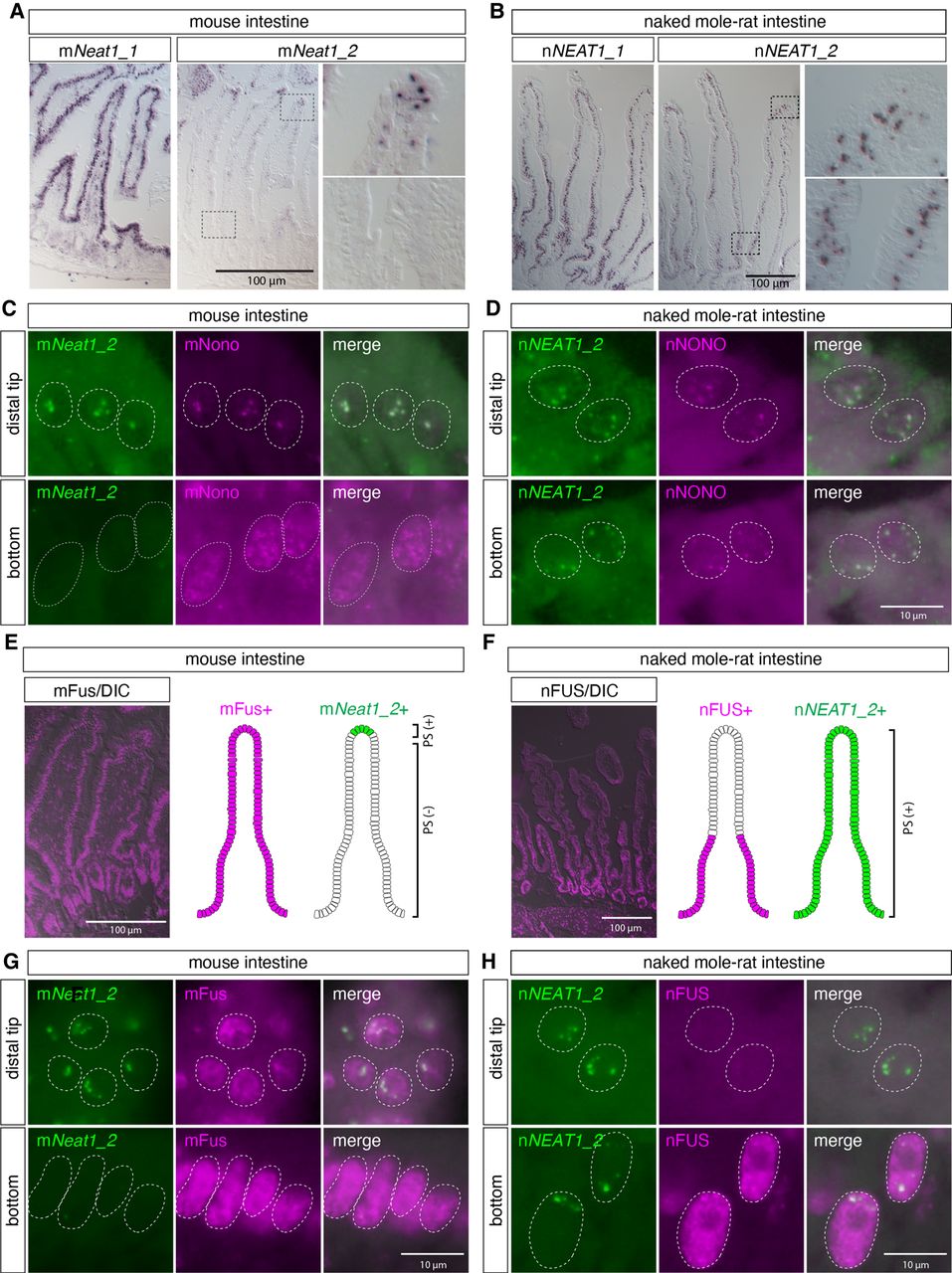
nNEAT1_2 is expressed in the entire regions of intestinal epithelium in naked mole-rats, whereas the essential paraspeckle component nFUS is not expressed in the distal region of intestinal epithelium in naked mole-rats. (A,B) In situ hybridization analyses of mNeat1/nNEAT1 in intestine of mice (A) and naked mole-rats (B). Note that the expression of mNeat1_2 is restricted to the distal tip cells in the intestinal epithelium of mice, whereas the expression of nNEAT1_2 is broadly observed throughout the epithelium in naked mole-rats. (C,D) Simultaneous detection of mNeat1_2/nNEAT1_2 (green) and the paraspeckle marker mNono/nNONO (magenta) in the top and the bottom regions of intestinal epithelium in mice (C) and naked mole-rats (D). Paraspeckle formation was observed in the bottom region of intestinal epithelium in naked mole-rats. (E,F) The expression of mFus/nFUS (magenta) in intestinal epithelium in mice (E) and naked mole-rats (F) and schematic drawing of the expression of mFus/nFUS and mNeat1_2/nNEAT1_2 (green) in the epithelium. DIC: differential interference contrast images. (G,H) Simultaneous detection mNeat1_2/nNEAT1_2 (green), and mFus/nFUS (magenta) in the top and the bottom regions of intestinal epithelium in mice (G) and naked mole-rats (H). Note the formation of paraspeckles in top region of the epithelium of naked mole-rats despite the lack of the expression of essential paraspeckle protein nFUS. Scale bars, 100 µm in A, B, E, F and 10 µm in C, D, G, H.




