


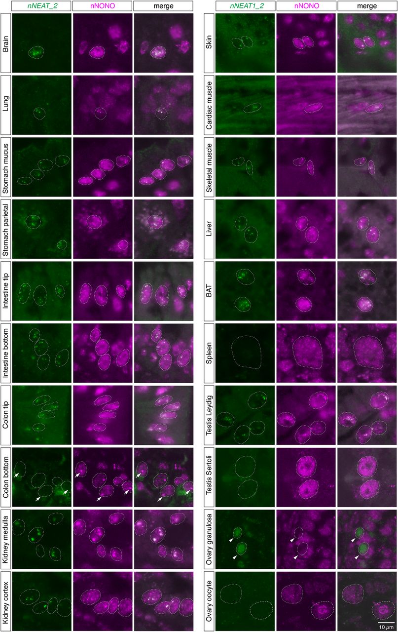
FIGURE 5.
Formation of paraspeckles in various cell types in naked mole-rat tissues. Simultaneous detection of nNEAT1_2 and a paraspeckle marker nNONO in various tissues of naked mole-rats. Prominent paraspeckle formations identified by the colocalization of nNEAT1_2 and nNONO were observed in a wide range of cell types. Arrows indicate the large nNONO foci in the bottom regions of colon epithelium that lacked the expression of nNEAT1_2. Arrowheads indicate a subset of granulosa cells that lacked the expression of nNONO, where nNEAT1_2 was diffusedly localized in the nucleus. Scale bar, 10 µm.




