


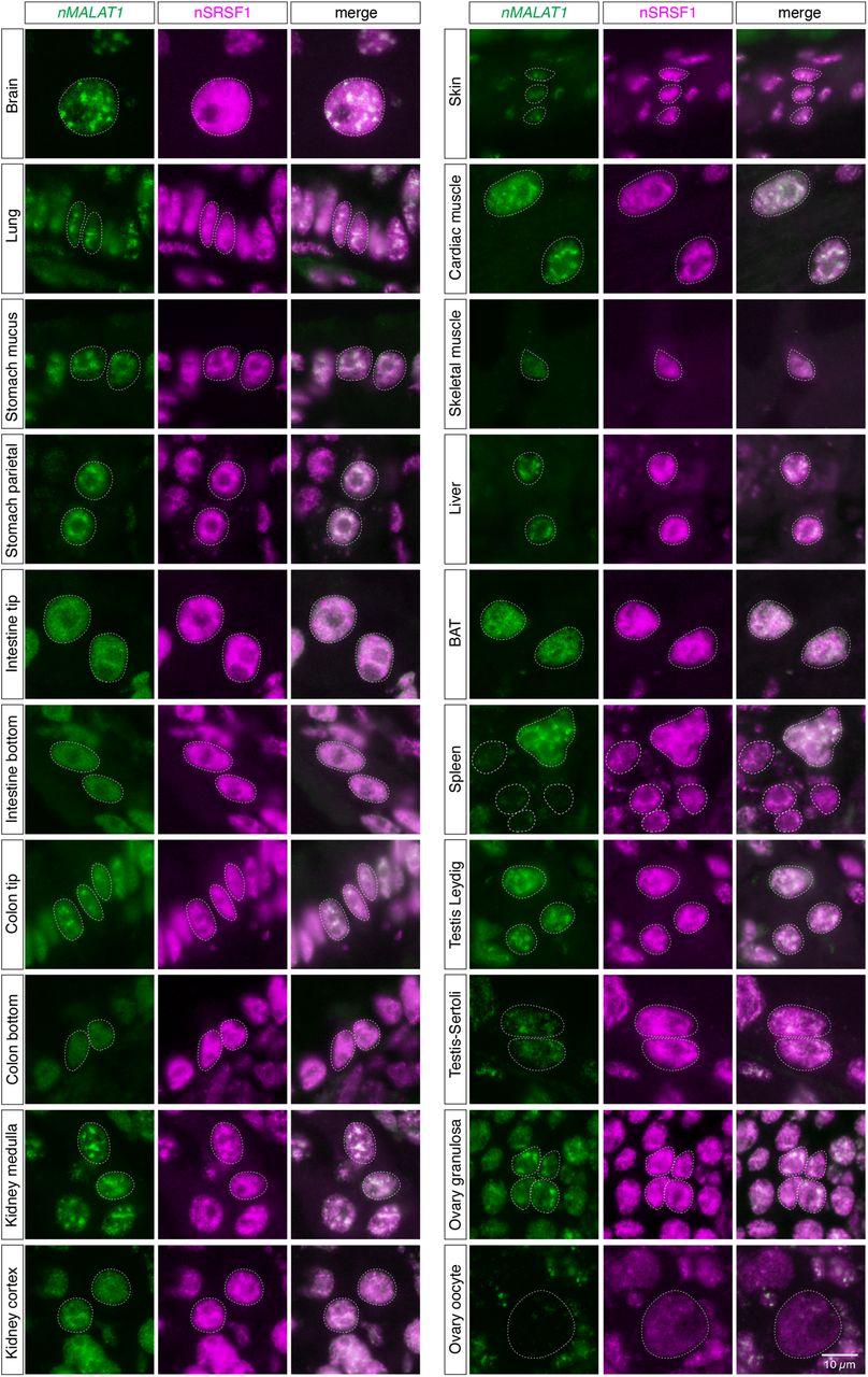
FIGURE 4.
Subnuclear localization of nMALAT1 exhibit various patterns in different cell types. Simultaneous detection of nMALAT1 and a nuclear speckle marker nSRSF1 in various tissues of naked mole-rats. Note that nMALAT1 was diffusedly distributed in the bottom regions of intestinal and colon epithelium and skeletal muscle cells, despite the speckled distribution of SRSF1. Scale bar, 10 µm.




