


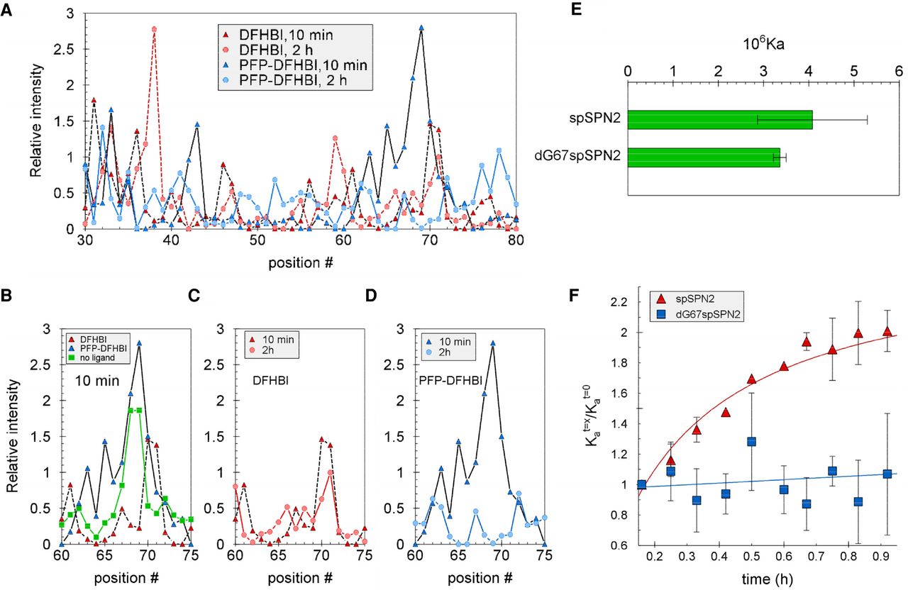
Evidence for an initial disruption by PFP-DFHBI around the SPN2 binding pocket entrance followed by structural uniformity around the pocket requiring the 2′OH on G67. (A–D) SPN2 was analyzed by SHAPE after 10- and 120-min incubations with DFHBI and PFP-DFHBI in buffer SX. (A) Relative intensities at all aptamer positions from incubations for 10 or 120 min with DFHBI or PFP-DFHBI. (B–D) Relative intensities in the region around the gateway A after incubation for (B) 10 min with DFHBI or PFP-DFHBI, (C) 10 or 120 min with DFHBI, or (D) 10 or 120 min and PFP-DFHBI. (E) The affinities for PFP-DFHBI of spSPN2 and dG67spSPN2 at 10 min. (F) Time course of the change in affinity for PFP-DFHBI of SPN2 and SPN2 with dG at position 67 (dG67SPN2).




