


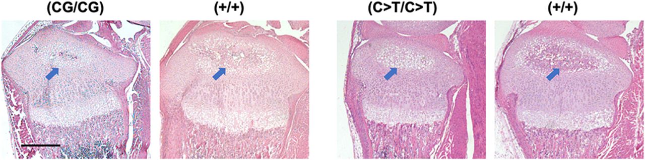
Histology of the proximal tibia of miR140-CG and -C > T models. Representative images of Hematoxylin and Eosin-stained proximal tibias of miR140-CG mice and miR140-C > T mice and control littermates at postnatal day 10. Homozygous miR140-CG [(CG/CG)] and -C > T [(C > T/C > T)] tibias present a modest delay in the maturation of chondrocytes with delayed development of the secondary ossification center (blue arrows) compared with wild-type littermate control [(+/+)]; in both CG and C > T bones, the secondary ossification is mostly occupied by hypertrophic chondrocytes at this stage (stained white due to a smaller amount of extracellular matrix), whereas in control wild-type bones, eosinophilic staining appears in the secondary ossification center, indicating replacement of hypertrophic chondrocytes by the bone matrix. Scale bar represents 500 µm.




