


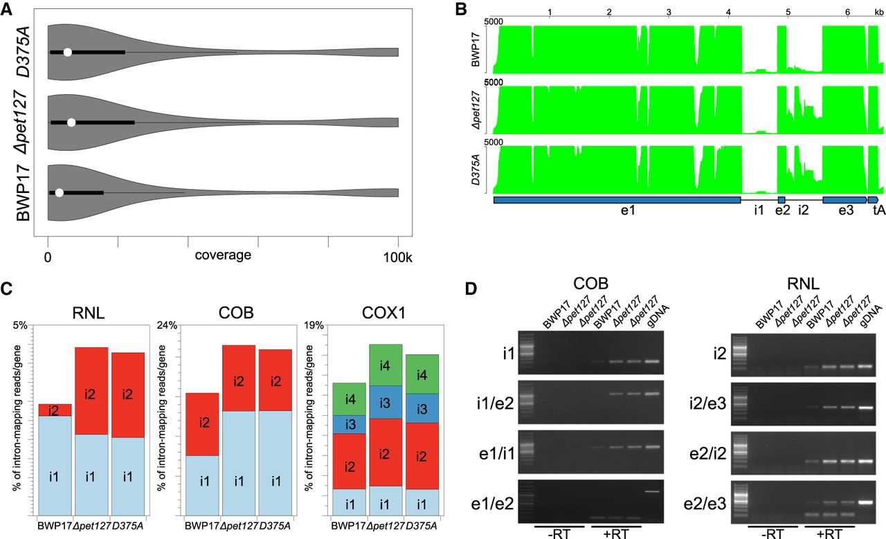
Increased intron accumulation in ΔCapet127 and Capet127D375A strains. (A) The distribution of sites in the mtDNA reference sequence with varying coverage in the wild-type (BWP17), homozygous ΔCapet127 mutant, and Capet127D375A catalytic mutant. Width of the plot corresponds to the frequency of sites covered by the number of RNA-seq reads shown on the x-axis. White circles and dark bars correspond to the median and the interquartile range (IQR), respectively. Coverage depth was calculated using the -depth option of SAMtools (Li et al. 2009) and visualized in R using the vioplot package. (B) Coverage by RNA-seq reads of the fragment of the C. albicans mtDNA reference sequence encompassing the RNL gene and the downstream tRNA-Ala [tA(UGC)mt] in the wild-type (BWP17), homozygous ΔCapet127 deletant, and Capet127D375A catalytic mutant. BWA files obtained using bamCompare (Ramírez et al. 2016) were visualized in pyGenomeTracks (Ramírez et al. 2018). The depth coverage axis was set at the maximum value of 5000 reads to better visualize low-coverage regions, truncating the highest values. (C) RNA-seq reads mapping to the intronic sequences in the RNL, COB, and COX1 genes, expressed as % of all reads mapping to the gene sequence (exons + introns). (D) Semiquantitative RT-PCR analysis of the introns and intron–exon junctions in the homozygous ΔCapet127 mutant (two independent repeats) compared to the wild-type (BWP17) strain. Fragments internal to the intron, encompassing the intron–exon junctions, and across introns were amplified following reverse transcription (+RT) of DNase-treated mitochondrial RNA. Amplification of reactions with the reverse transcriptase omitted (−RT) were used to control for DNA contamination of the RNA samples, and genomic DNA (gDNA) was used as a positive control.




