


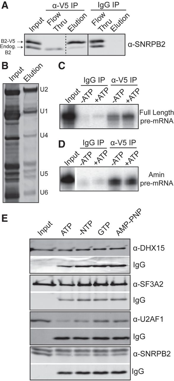
ATP hydrolysis alters U2 snRNP protein composition. (A) Western blot analysis of IP with anti-V5 and mouse IgG from HeLa nuclear extract containing V5-tagged SNRPB2 and probed with anti-SNRPB2, which detects both endogenous (endog) and V5-tagged SNRPB2 (V5-B2). (B) Denaturing gel analysis of RNA isolated from anti-V5 IP eluate stained with SYBR Gold. (C) Denaturing gel analysis of RNA isolated anti-V5 IP of in vitro splicing reactions ±ATP using a radiolabeled full-length RNA substrate in the conditions. (D) Same as C, except with an Amin substrate. (E) Western analysis anti-V5 of IP's from nuclear extract incubated under in vitro splicing conditions in the presence of ATP, GTP, or AMP–PNP. Blots were probed with the indicated antibody. Bands labeled with IgG show the amount of anti-V5 antibody in the elutions.




