


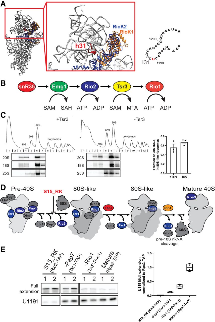
Tsr3 functions in 80S-like ribosomes. (A) (Left) Superimposition of two human pre-40S structures (PDBID 6ZXE and 6G18, respectively) based on Rps15 (uS19) shows overlap of human Rio1 (RioK1, colored in orange) and human Rio2 (RioK2, colored in blue). 18S rRNA from Rio1 containing pre-40S (PDBID 6ZXE) is shown in gray ribbon, with h31 highlighted in red and U1248 (U1191 in yeast) highlighted in red sphere. (Right) Secondary structure of 18S loop 31 (l31), which is bound by all factors during assembly and contains the snR35, Emg1, and Tsr3 modification site at U1191. (B) snR35, Emg1, Rio2, Tsr3, and Rio1 all bind loop 31 at successive stages of 40S maturation. Emg1 and Tsr1 use S-adenosyl-methionine (SAM) to produce S-adenosyl-homocysteine (SAH) or methylthioadenosine (MTA), respectively, while Rio2 and Rio1 hydrolyze ATP. (C) Absorbance profiles at 254 nm and corresponding northern blots of 10%–50% sucrose gradients from Gal:Fap7 (+Tsr3) or ΔTsr3, Gal:Fap7 (−Tsr3) cells. In all cases, Fap7 was depleted in glucose for over 16 h to accumulate 80S-like ribosomes. Fractions 2–7 for each gradient were probed for 20S, 18S, and 25S rRNA. Quantifications of the data on the left indicate the efficiency of formation of 80S-like ribosomes. Data are shown as mean with standard deviation (n = 3). (D) Simplified scheme showing ordered release of Ltv1, Enp1, and Rio2 (blocked by Rps15_RK) before formation of 80S-like ribosomes and different 80S-like ribosomes that accumulate in the absence of Fap7 and Rio1, respectively. Tagged proteins for pulldowns are indicated in blue. Note that Rps3 is bound in all the stages depicted here, but because mature 40S are so much more abundant the majority of Rps3-TAP-tagged subunits are mature. (E) (Left) Primer extension results indicating the installation of acp modification at U1191 in different purified pre-40S intermediates and mature 40S. Signal for U1191 was first normalized to full extension before further normalization to mature 40S (Rps3-TAP). Two representative biological replicates (1, 2) are shown for each cell type. (Right) Quantification of the normalized U1191 stop from the left and two additional replicates (n = 4). Endogenous Rps15, Fap7, and Rio1 were depleted by growth in glucose of cells containing the respective proteins under the GAL-promoter. In the case of Rps15_RK, these cells were supplemented with a plasmid encoding Rps15_RK (Huang et al. 2020).




