


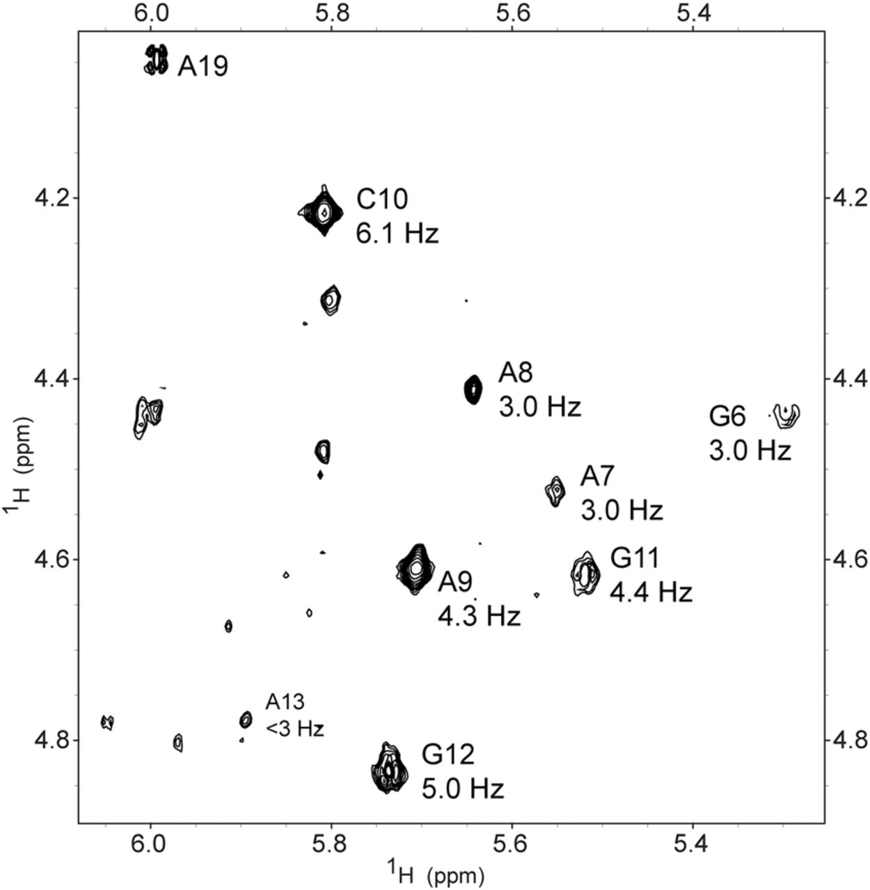
FIGURE 4.
NMR 2D TOCSY spectrum of 3 mM HP1 at 25°C with mixing time of 29 msec in D2O. H1′–H2′ cross-peaks are labeled with corresponding residue. The magnitude of the H1′–H2′ scalar-coupling constant (3J) determined from splitting of H1′ peaks in a 400 msec mixing time NOESY spectrum is given below the residue identifier. Coupling accuracy is approximately ±0.5 Hz.




