


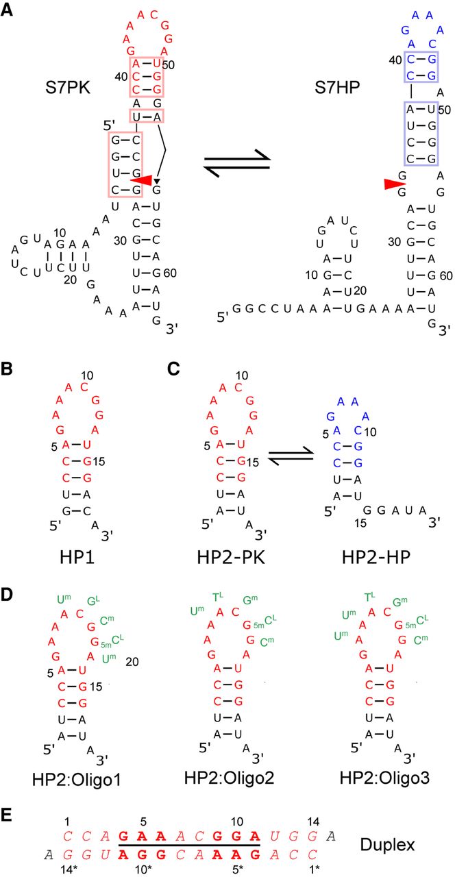
(A) Conserved region of influenza A segment 7 mRNA showing the 3′ splice site (red triangle) in two secondary structures predicted from sequence comparison (Table 1): a pseudoknot with hidden splice site (S7PK) and a long hairpin with exposed splice site (S7HP). Colored nucleotides are included in model hairpins shown in B–D. Colored boxes correlate with colors highlighting base pairs in Table 1. (B,C) Two 19-nt hairpins, HP1 and HP2, used for NMR studies. HP1 (B) and HP2 (C) shown in two hairpin loop conformations corresponding to those predicted in pseudoknot (HP2-PK) and hairpin (HP2-HP) of segment 7 mRNA. (D) HP2 with Oligonucleotides 1, 2, and 3 designed to target the loop as shown. The 5′ end of each oligonucleotide is assigned number 20. In the oligonucleotide residue names, “L” is LNA, “m” is 2′-O-methyl in the sugar, and “5m” is 5-methyl in the base. TL is thymidine LNA. (E) Self-complementary duplex with 8 × 8-nt internal loop formed by 5′CCAGAAACGGAUGGA at 100 mM K+ (Kauffmann et al. 2017).




