


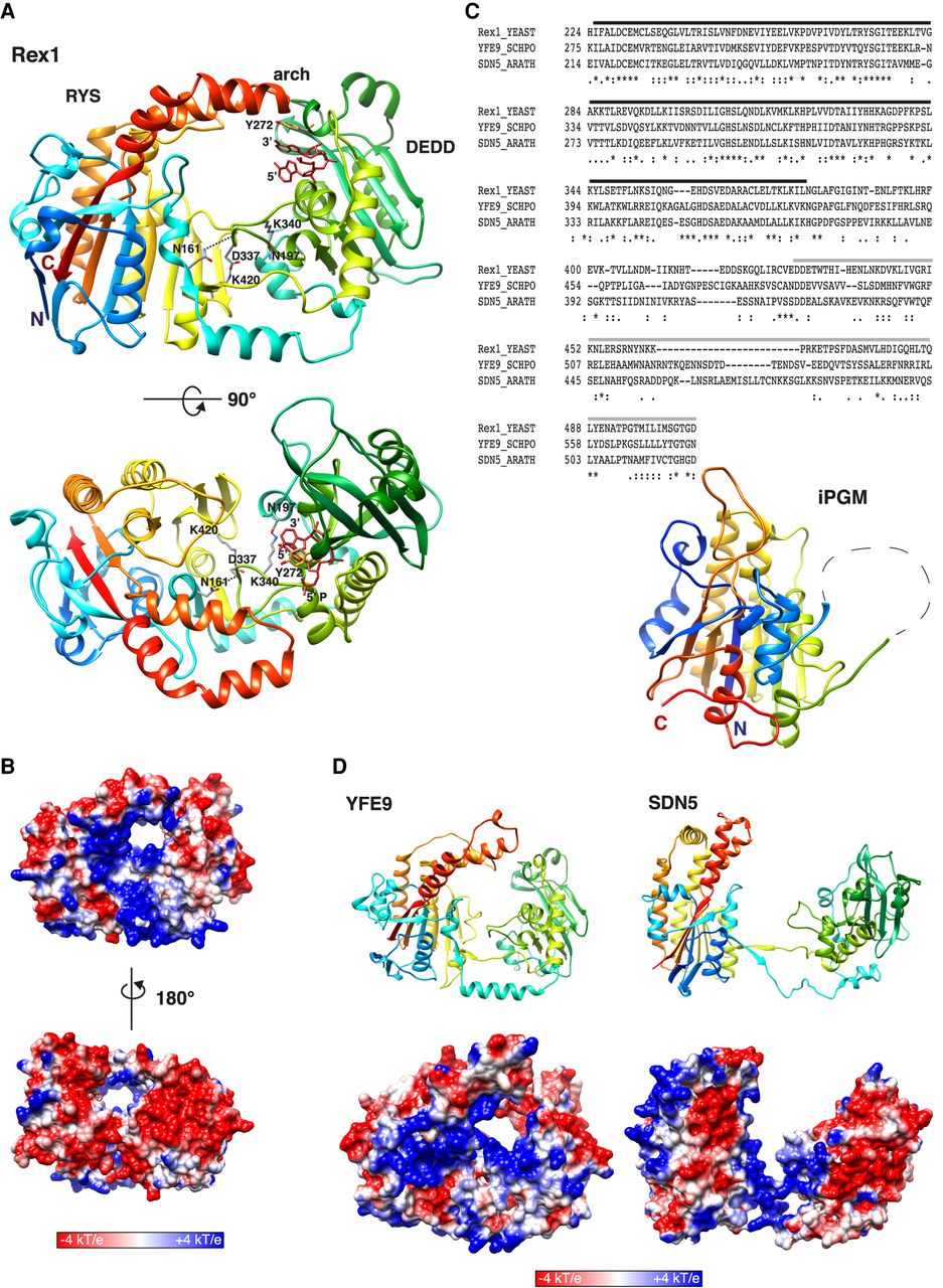
The Dual domain structure of Rex1. (A) Ribbon structure of the AlphaFold model of Rex1 (residues 53–553) with bound substrate. The terminal dinucleotide from the threaded Rex1/RNA model (Fig. 1E) was superposed onto the Rex1 AlphaFold model by manual alignment with residue Y272. The Y272 side chain is shown in both models (gray and gold) to indicate the alignment. Front and top views are shown. Coloring is from blue (amino terminus) to red (carboxyl terminus). The DEDD domain, the RYS domain and the helical arch are labeled. Interactions between specified residues are shown. The 3′ and 5′ nt are labeled. The 5′ phosphate group is labeled in the lower panel. (B) Electrostatic potential map of Rex1. Front and rear views are shown. (C) BLAST sequence alignment of Rex1, YFE9, and SDN5. The DEDD domain is overscored in black. iPGM- and PPM-related sequences are overscored in gray. The fold of the substrate binding domain of iPGM from B. stearothermophilus (residues 2–78 and 309–511) is shown. (D) Ribbon structures and electrostatic potential maps of (left panels) YFE9 (residues 87–631) and (right panels) SDN5 (residues 41–567). Amino-terminal sequences of the Rex1, YFE9, and SDN5 AlphaFold models with low confidence scores are excluded. Orientations of iPGM, YFE9, and SDN5 are shown as for Rex1 in A.




