


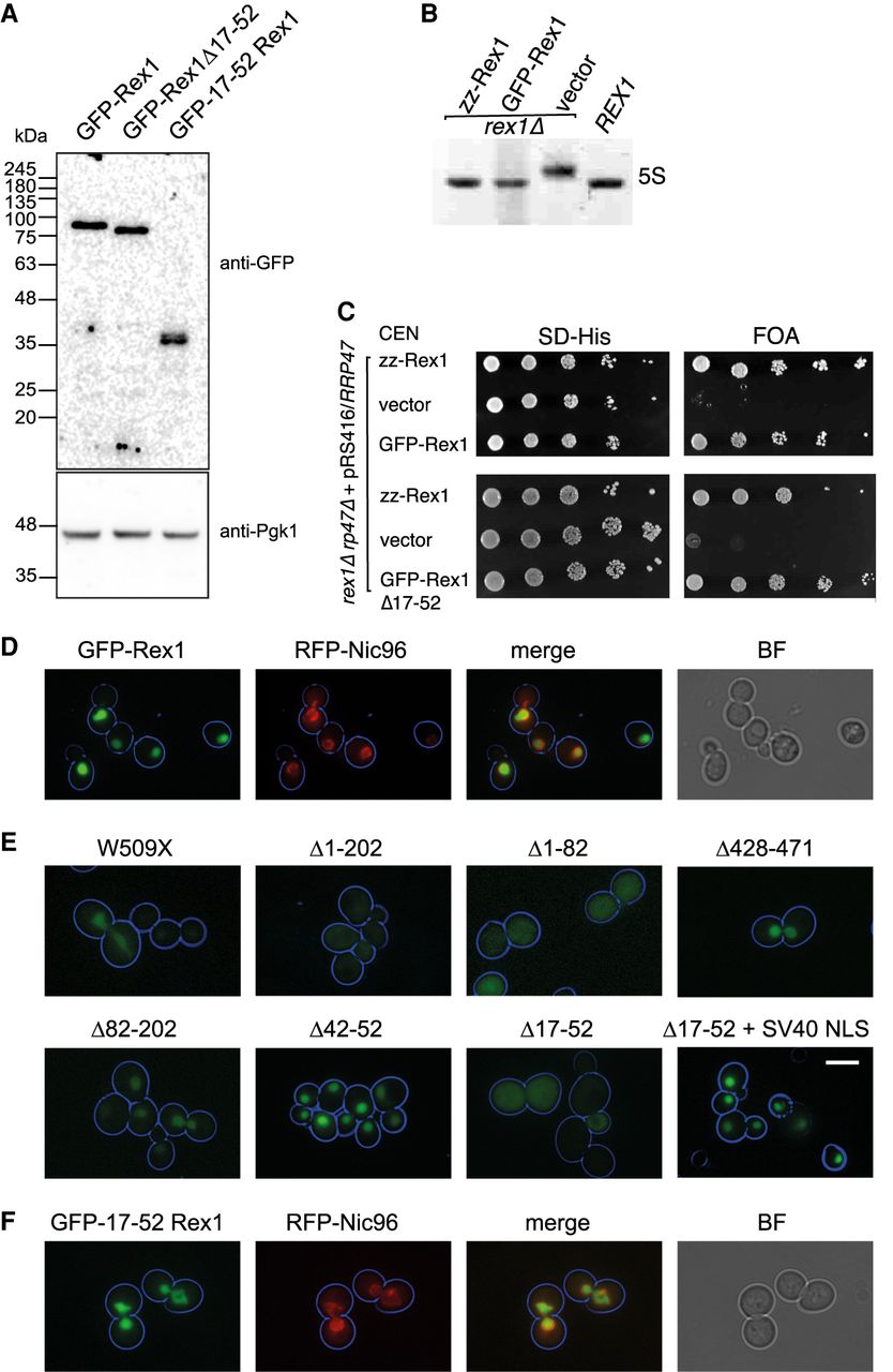
Identification of a nuclear localization signal in Rex1. (A) Western analysis of whole-cell extracts from a rex1Δ mutant expressing GFP–Rex1 fusion proteins. (B) Ethidium-stained RNA gel showing the relative migration of 5S rRNA species from a REX1 wild-type strain and rex1Δ mutants expressing either Rex1 zz or GFP fusions. (C) Plasmid shuffle assays on rex1Δ rrp47Δ double mutants expressing Rex1 GFP and zz fusion proteins. (D–F) Epifluorescence microscopy images of cells expressing versions of GFP–Rex1 and mRFP–Nic96. (D) Colocalization of GFP–Rex1 and mRFP–Nic96. (E) Localization of GFP–Rex1 deletion mutants. (F) Colocalization of GFP fused to residues 17–52 of Rex1 and mRFP–Nic96. Cell circumferences are indicated in blue. Bar, 5 µm. (BF) Brightfield, (SV40-NLS) simian vacuolating virus 40 nuclear localization signal.




