


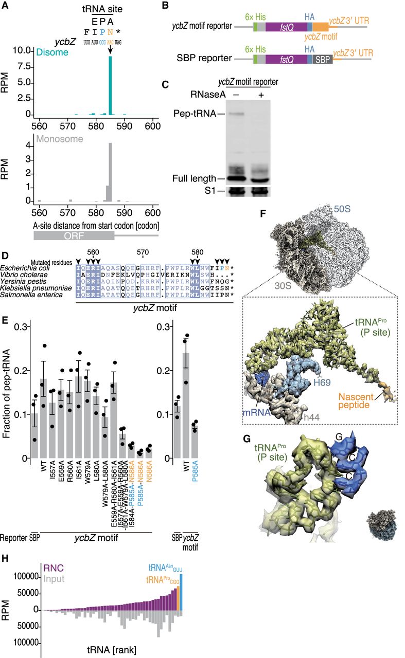
Ribosomes stall on ycbZ mRNA. (A) The footprint distribution along ycbZ in disome profiling (top) and monosome profiling (bottom). The A-site position (in the case of disome, A-site of leading ribosome) of the reads is shown. The identified pause site is indicated by an arrow. (B) Schematic representation of reporter mRNAs used in this study. (C) Western blot analysis of the translation product (probed by HA epitope) from the ycbZ motif reporter subjected to SDS-PAGE at neutral pH. RNase treatment verified the RNA conjugation. (Pep-tRNA) Polypeptidyl-tRNA. (D) Conservation of the ycbZ pause motif. The amino acid position in E. coli is shown. Mutated amino acids (tested in E) are indicated by arrowheads. (E) Mutational analysis of the ycbZ motif. The same experiments as in C were performed with ycbZ motif and SBP reporters. The quantification (mean and SD) from three replicates is shown. (F,G) Cryo-EM structure of the ycbZ-stalled ribosomes. Close-up view of the intersubunit cavity of this complex depicting the structural details of the nascent peptide-bound P-site tRNA (F). The structural model of tRNAProCGG is well fitted to the corresponding cryo-EM density. The details of the codon–anticodon recognition of P-site tRNA are shown in G. (H) Enrichment of tRNAs in the RNC formed on the ycbZ motif, as assessed by tRNA-seq. (RPM) Reads per million mapped reads.




