


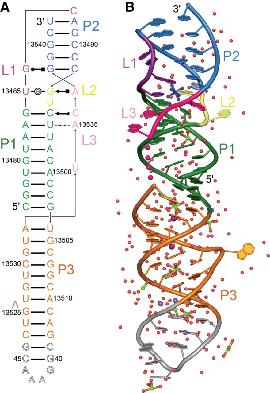
Structure of the SARS-CoV-2 frameshifting pseudoknot. (A) Secondary structure. Arrows denote chain connectivity; Leontis and Westhof (2001) symbols, non-Watson–Crick base pairs. Numbering corresponds to the SARS-CoV-2 reference genome (NC_045512.2), except for residues 39–46 (outlined letters) introduced to facilitate crystallization. (B) Cartoon representation of the 1.3 Å-resolution structure of the pseudoknot, colored as in A. Nucleotides altered for crystallization are gray. Pink spheres denote chain connectivity in lieu of U13534, which is disordered. For alternate conformers, bases are in pastel shades. Red, lime, light purple, and purple spheres represent water molecules, Mg2+, Na+, and K+, respectively. Cobalt hexammine is colored blue.




