


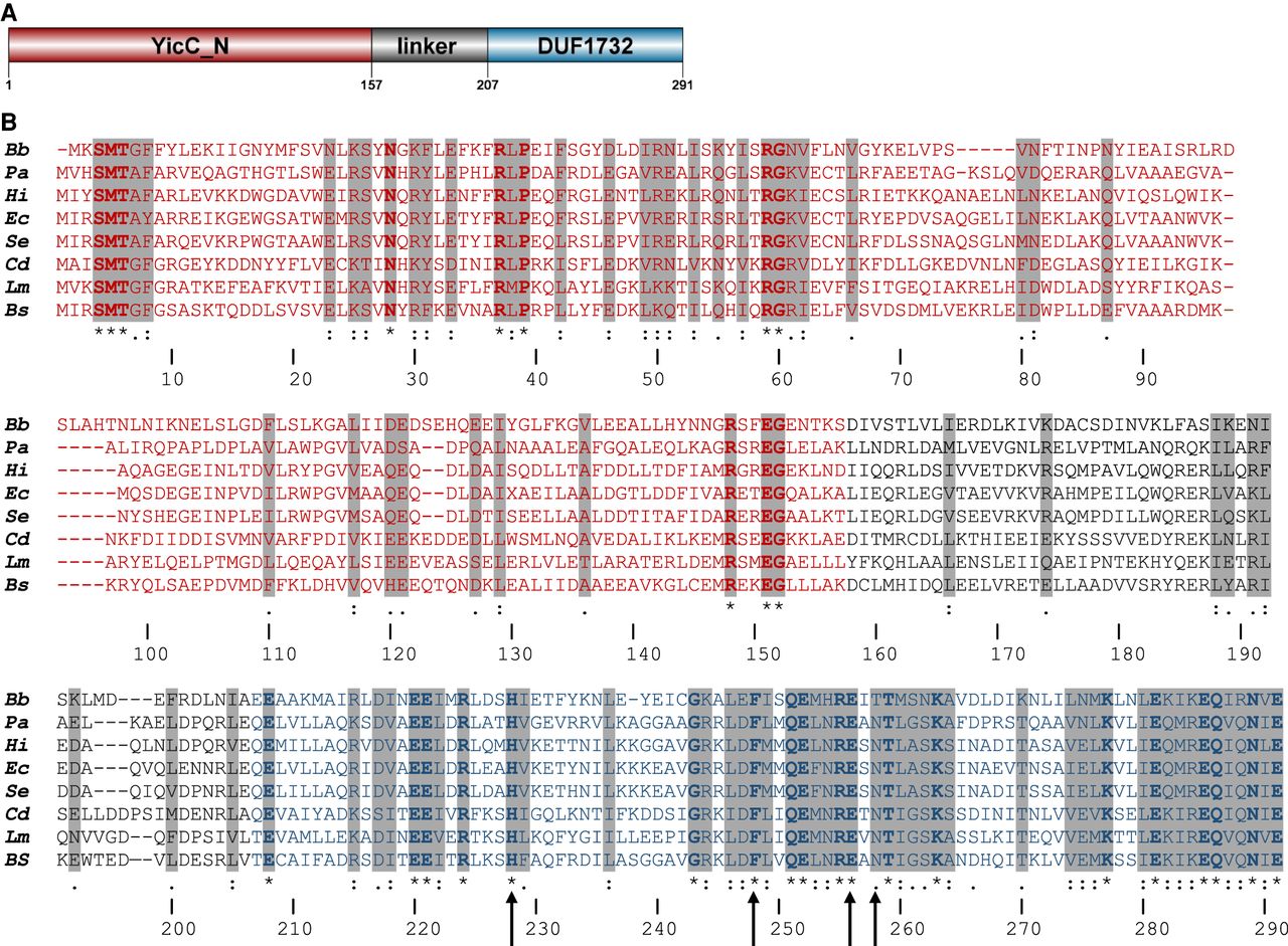
YloC domain structure and primary sequence. (A) Domain structure of YloC, as predicted by the Pfam database of protein families (Mistry et al. 2021). (B) CLUSTAL alignment of YloC-like proteins from eight organisms. Amino acid residues in domains are colored as in the schematic in part A. Two-letter abbreviations: (Bb) Borrelia burgdorferi, (Pa) Pseudomonas aeruginosa, (Hi) Haemophilus influenza, (Ec) Escherichia coli, (Se) Salmonella enterica, (Cd) Clostridia difficile, (Lm) Listeria monocytogenes, (Bs) Bacillus subtilis. Upward-pointing arrows below the alignment point to residues (H228, F248, E256, and N258) that were mutated to alanine. Asterisks indicate identical residues in all species. Two dots indicate conserved residues. One dot indicates similar residues. Regions of high homology are shaded.




