


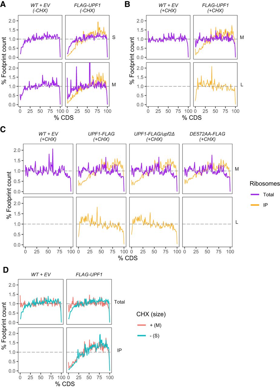
Upf1 progressively associates with ribosomes across mRNA coding regions except when Upf1 association forms L footprints. Distribution of footprint abundance across the coding region (CDS) for each footprint size class from total (purple) or IP (orange) ribosomes profiling libraries. (A) Small (S) and medium (M) footprint distribution from strains expressing WT + EV and FLAG-UPF1 in the absence of CHX treatment. (B) Medium (M) and large (L) footprint distribution from strains expressing WT + EV and FLAG-UPF1 in the presence of CHX. (C) Medium (M) and large (L) footprint distribution from CHX-treated cells expressing WT + EV, UPF1-FLAG, UPF1-FLAG/upf2Δ, or upf1DE572AA-FLAG. Each gene's CDS was divided into 100 bins and the percentage of footprints’ P-sites belonging to each bin in all genes was calculated. Gray dashed line signifies a theoretical number where each percentage of CDS contains an equivalent number of footprints for a sum of 100% across all 100 bins. (D) Distribution of dominant footprint (red = Medium, M for +CHX; green = Small, S for −CHX) abundance across the coding region. Data is the same as used in A and B top panels but replotted in such a way to directly observe the differences or similarity in footprint distribution between the absence and presence of CHX during the library preparation of each strain.




