


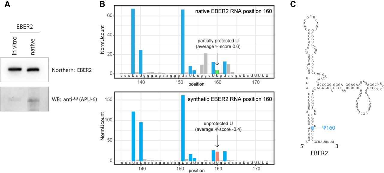
EBER2 is pseudouridylated at U160. (A) Antibody-mediated detection of pseudouridylation in EBER2. Native EBER2, purified from EBV-infected cells, as well as in vitro-transcribed EBER2, was probed with an anti-Ψ antibody (monoclonal antibody APU-6) to detect the presence of pseudouridine and verify our mass spectrometry results. Northern blot analysis for EBER2 was carried out with the same blot to verify comparable loading. (B) HydraPsiSeq analysis maps the pseudouridylation site to U160 of EBER2. HydraPsiSeq was carried out in parallel with native and in vitro-transcribed EBER2. Protected U160 is shown by an arrow. Gray bars indicate cleavage at non-U residues. (C) Secondary structure of EBER2; the pseudouridylation site Ψ160 is highlighted.




