


FIGURE 3.
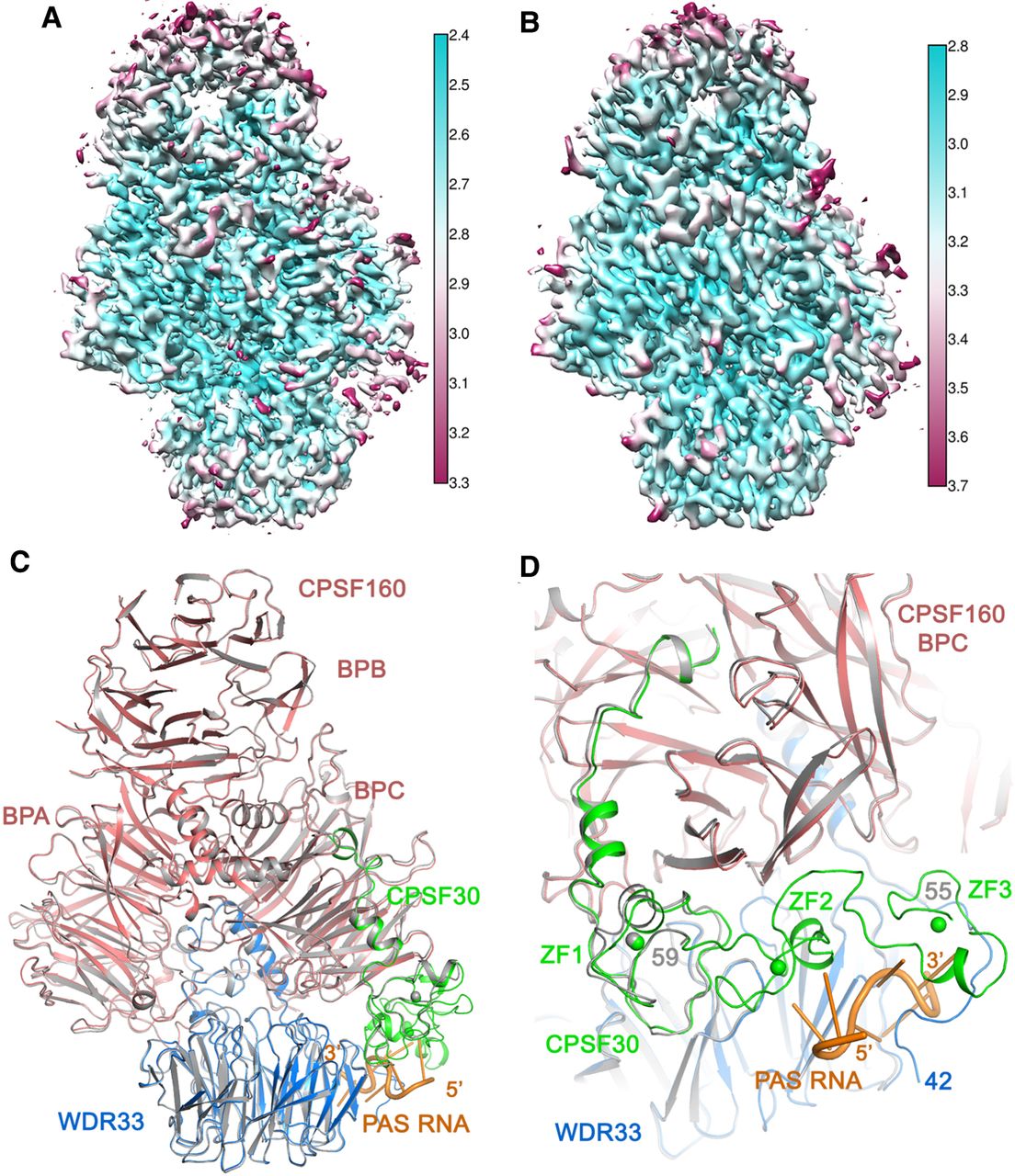
Overall structure of the human CPSF160–WDR33–CPSF30 ternary complex. (A) Local resolution map for the sample containing AAGAAA RNA. The lack of RNA binding is visible by comparing the lower right corner with Figure 1C. (B) Local resolution map for the sample containing GAUAAA RNA. (C) Overlay of the structure for the AAGAAA sample (gray, lacking RNA) with that of the AUUAAA quaternary complex (in color). (D) Disordering of ZF2 and ZF3 of CPSF30 and residues 42–54 in the amino-terminal extension to the WD40 domain of WDR33 in the AAGAAA sample (in gray) compared to the AUUAAA quaternary complex (in color). Panels A and B were produced with Chimera.




