


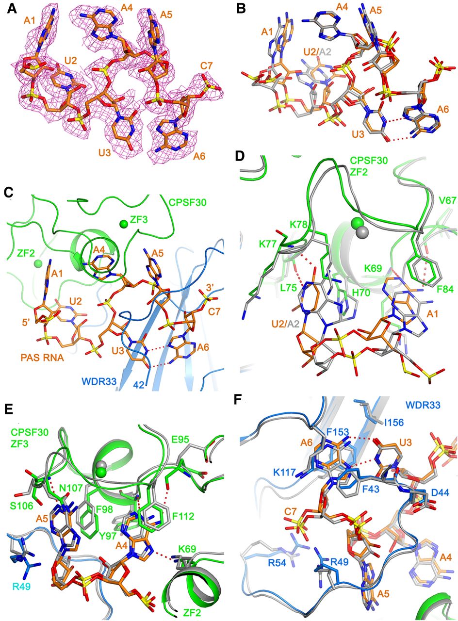
Recognition of the AUUAAA PAS RNA. (A) Cryo-EM density (magenta mesh) for AUUAAA of the PAS RNA (orange), as well as the 5′ phosphate of the nucleotide (C7) immediately following the PAS. (B) Overlay of the binding mode of the AUUAAA PAS (orange) and the AAUAAA PAS (gray). Hydrogen bonds in the U3–A6 Hoogsteen base pair are indicated with dashed lines in red. (C) Binding mode of the AUUAAA PAS (orange) at the interface of CPSF30 ZF2-ZF3 (green) and WDR33 (light blue). (D) Detailed interactions showing the recognition of the A1 and U2 bases of the PAS (orange) by ZF2 of CPSF30 (green). Hydrogen-bonds between the adenine bases and the protein are indicated with dashed lines in red. The binding of the A1 and A2 bases in the AAUAAA complex is shown in gray. (E) Detailed interactions showing the recognition of the A4 and A5 bases of the AUUAAA PAS (orange) by ZF3 of CPSF30 (green). (F) Recognition of the U3–A6 base pair in the AUUAAA PAS (orange) by WDR33 (blue).




