


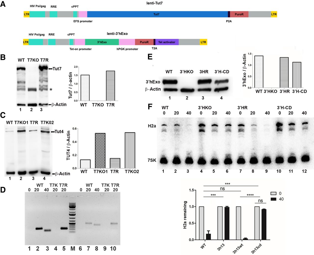
Rescue of the TUT7 and 3′hExo KO cells. (A) Schematic of lentiviral vectors used for expression of TUT 7 (top) and 3′hExo (bottom). The 3′hExo vector is TET inducible, and the TUT7 vector is not (since the size of the TUT7 ORF was too large to also include the TET regulatory system). (B) TUT7 KO cells were transduced with the lentiviral vector expressing TUT7. Clonal cell lines were isolated from puromycin resistant cells. A western blot WT (lane 1), T7KO1 (lane 2), and of a cell line expressing similar amounts of TUT7 as the WT cells in shown (lane 3). The (*) indicates a cross-reacting band. Other bands in the WT and rescued cells are likely proteolytic products of TUT7. (C) Both the TUT7KO1 and TUT7KO2 cells were rescued with the lentiviral vector. The levels of TUT4 protein in the KO and rescued cells were determined by western blotting. The levels of TUT4 are increased in the KO cells and decreased to levels similar to the WT in the rescued cells. (D) The TUT7 rescue cells rescue the uridylation of histone mRNA degradation. Uridylated histone mRNA degradation intermediates were detected by RT-PCR using a reverse primer that ended in 3A's and a forward primer in two different histone H2a genes, as described in Mullen and Marzluff (2008). RNA from exponentially growing cells (lanes 1 and 6) and cells treated for 20 (lanes 2 and 7) or 40 min with HU (lanes 3 and 8) was analyzed. RNA from the TUT7 KO cells (lanes 4 and 9) and the TUT7 rescued cells (lanes 5 and 10) after 20 min of HU treatment were analyzed to demonstrate the rescue of uridylation. (E) The 3′hExo KO cells were rescued using the lentiviral vector containing the TET-ON system expressing either the active or catalytically dead 3′hExo. Cloned cell lines were isolated and the levels of tetracycline necessary to restore close to normal expression of 3′hExo were determined (Supplemental Fig. S3A–D). Western blots showing 3′hExo levels in the WT, KO, and cells rescued with WT 3′hExo (3HR) or catalytically dead (3′H-CD) 3′hExo relative to the levels of actin are shown. (F) The levels of histone mRNA in the WT (lanes 1–3), 3′hExoKO (lanes 4–6), KO cells rescued with WT 3′hExo (lanes 7–9), and KO cells rescued with catalytically dead 3′hExo (lanes 10–12) before (lane 1,4,7,10) and after treatment with HU for 20 (lanes 2,5,8,11) or 40 (lanes 3,6,9,12) minutes. Histone H2a mRNA levels were determined by northern blotting using the 7SK RNA as an internal control. The results of three independent experiments were quantified using a PhosphorImager.




