


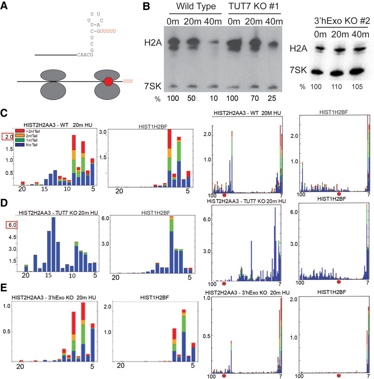
Effect of KO of TUT7 and 3′hExo on histone mRNA degradation. (A) Diagram of the two major classes of degradation intermediates in histone mRNA. The initial step is degradation 3′ to 5′ by 3′hExo into the stem where there is extensive uridylation of the intermediates. A second phase is rapid degradation until the exosome nears the terminating ribosome, resulting in an intermediate about 15 nt 3′ of the stop codon (red octagon). There is then slower degradation through the ORF of the mRNA with uridylation of many intermediates. (B) S-phase cells (3 h after release from double thymidine block) were treated with 5 mM hydroxyurea (HU) and RNA isolated from untreated cells, 20 and 40 min after treatment with HU. RNA was analyzed by northern blotting for the histone H2a mRNAs. 7SK RNA was measured as a loading control. The amount of histone mRNA was determined by PhosphorImager analysis with 7SK as the control. In the experiment with the 3′hExo KO, a different preparation of 7SK probe was used which had a higher specific activity than the probe used on the left panel for the TUT7 KO. (C–E) Libraries containing histone mRNAs were prepared from the WT, TUT7 KO, and 3′hExo KO cells 20 min after HU treatment, as previously described (Welch et al. 2015). The results for the 3′ end of two histone mRNAs, HIST2HAA3 which has a long 3′-UTR and HIST1H2BF which has a short 3′-UTR, are shown. The left panel shows the intermediates in the stem (nts 5–20), which were plotted showing the amount of uridylation of each intermediate as in Figure 2. The right panel plots the intermediates from 7–100. The position of the stop codon in each gene is shown by the red octagon. The y-axis shows the percentage of the intermediates relative to the total histone mRNA. Thus there are about 3× as many intermediates in the H2AA3 TUT7 library as in the wild-type library, and half as many intermediates in the 3′hExo library.




