


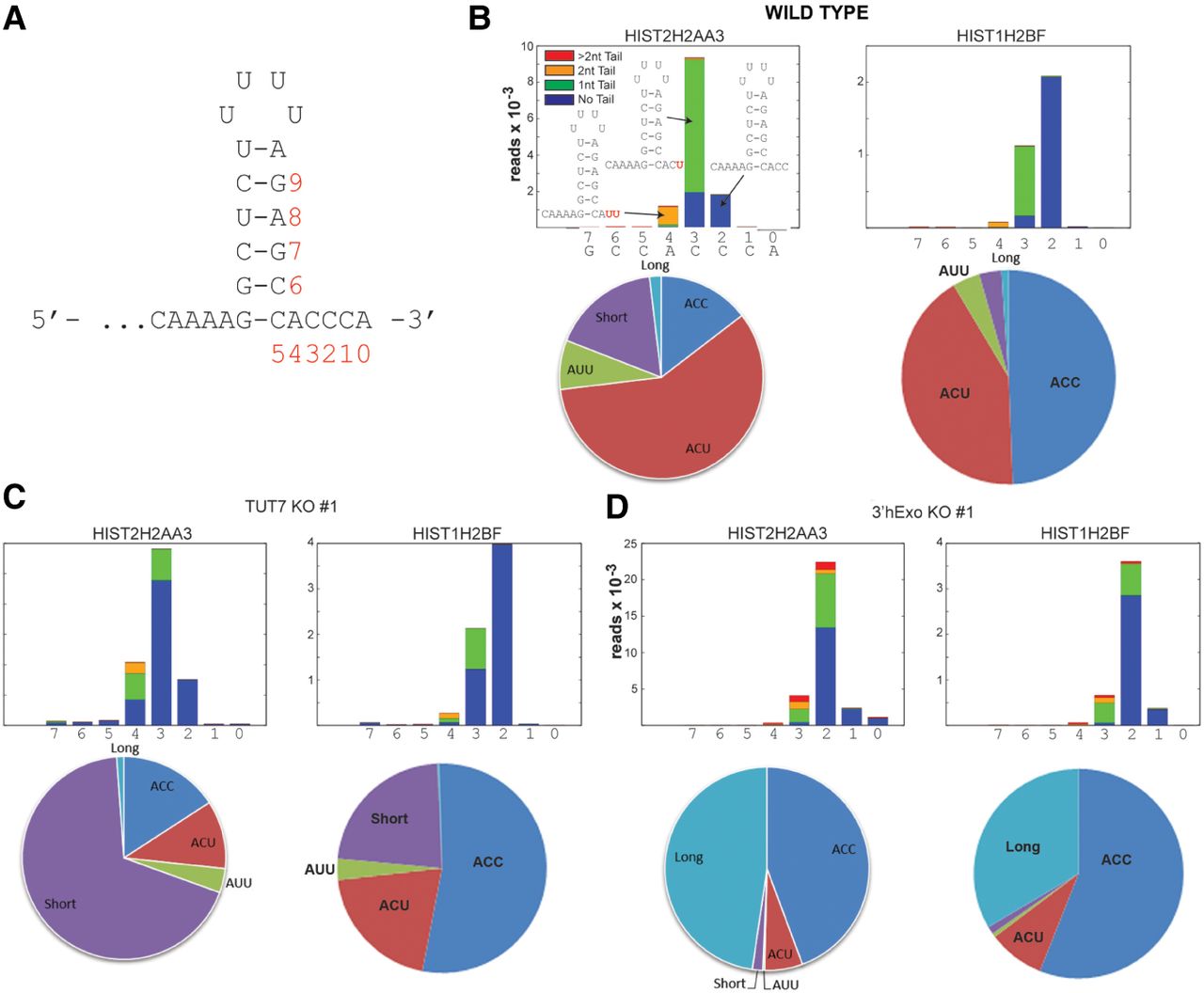
Analysis of the 3′ ends of the histone mRNAs in the KO cells. (A,B) The structure of the 3′ end of histone mRNA formed by 3′ processing in the nucleus. The 3′ ends of histone mRNAs were determined by the End-seq protocol with the results plotted as previously described (Welch et al. 2015). The position of the last templated nucleotide in the histone mRNA is indicated. 3′hExo removes 2 nt from the 3′ end of the processed histone mRNA. If more nucleotides are removed, a terminal uridyl transferase (TUTase) adds uridines back to the 3′ end with the number of uridines added indicated by the colored blocks. The structure of the three most abundant 3′ ends is shown. The pie chart shows the percentage of reads with the different 3′ ends in the HIST2HAA3 gene, with short and long indicating the amounts of the 3′ ends that were either shorter or longer than the major forms (ACC, ACU, and AUU) which extend 3 nt past the base of the stem. The results for two different histone mRNAs are shown. (C,D) The same analysis was performed for the TUT7 KO cells (C) and for the 3′hExo KO cells (D). The y-axis gives the number of reads for each index (×10−3).




