


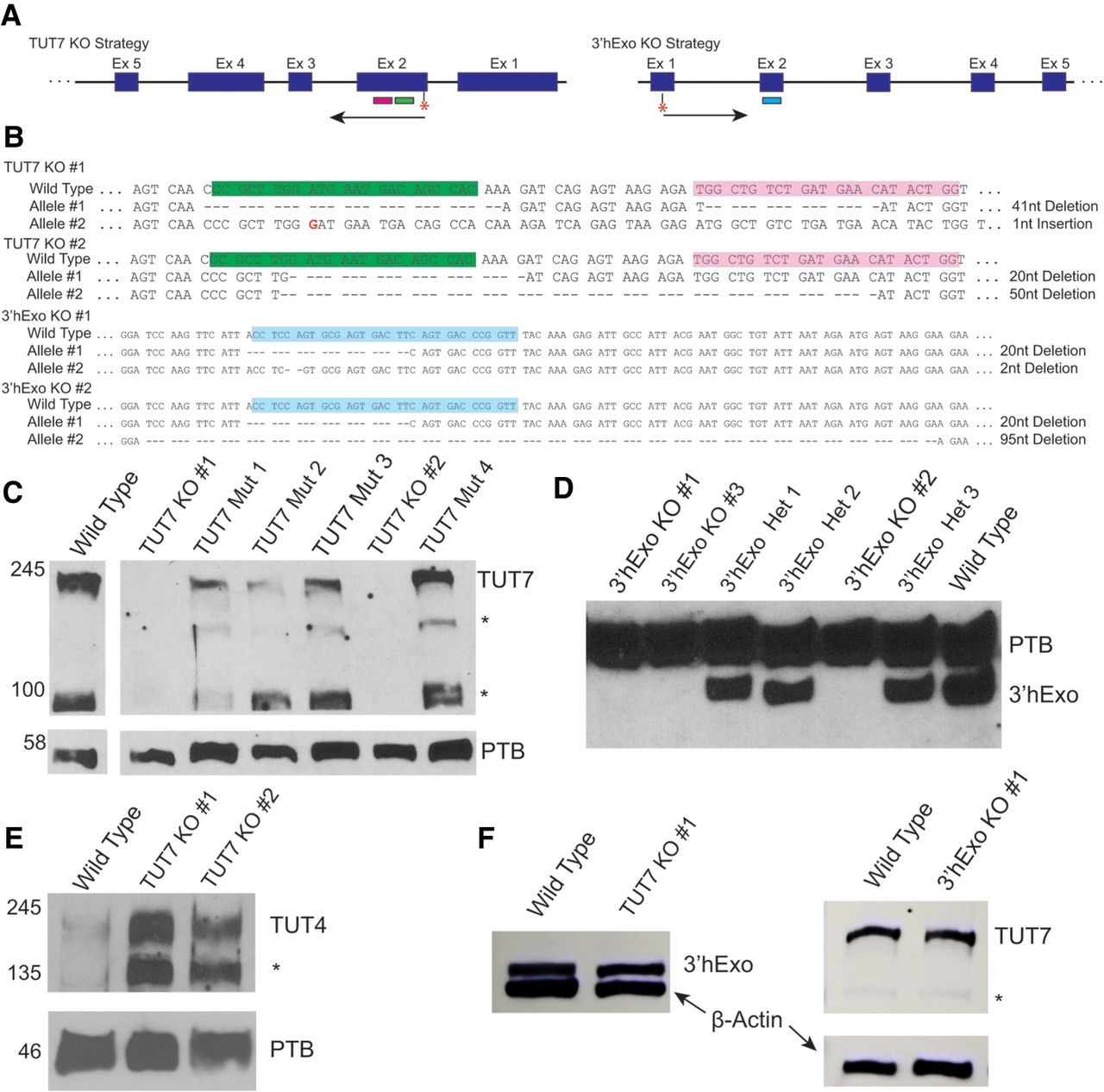
Characterization of 3′hExo and TUT7 KOs. (A) Structure of the TUT7 (left) and 3′hExo genes (right). The (*) indicates the start codon, and the colored boxes the location of the guide RNAs. (B) CRISPR strategy and results. The guide RNAs and NGG sequence are boxed. The two alleles for each KO were sequenced. Two independent TUT7 KO lines were isolated and several 3′hExo KO lines were isolated and two were characterized in detail. (C) Western blot analysis of potential TUT7 KOs. Six potential TUT7 KO lines were analyzed by western blotting. Four lines had only one allele inactivated and two had both alleles inactivated. (D) Western analysis of 3′hExo KOs. Three had both alleles inactivated and three were heterozygous, with a single allele inactivated. (E) The levels of TUT4 in the TUT7 KO cells were determined by western blotting. Each TUT7 KO had a substantial increase in expression of TUT4. (F) The levels of TUT7 and 3′hExo were determined in the KO of the other protein. The levels of TUT7 were similar to wild-type in the 3′hExo KO (right), and the levels of 3′hExo were similar to wild-type in the TUT7 KO (left). PTB or β-actin were analyzed as the loading control.




