


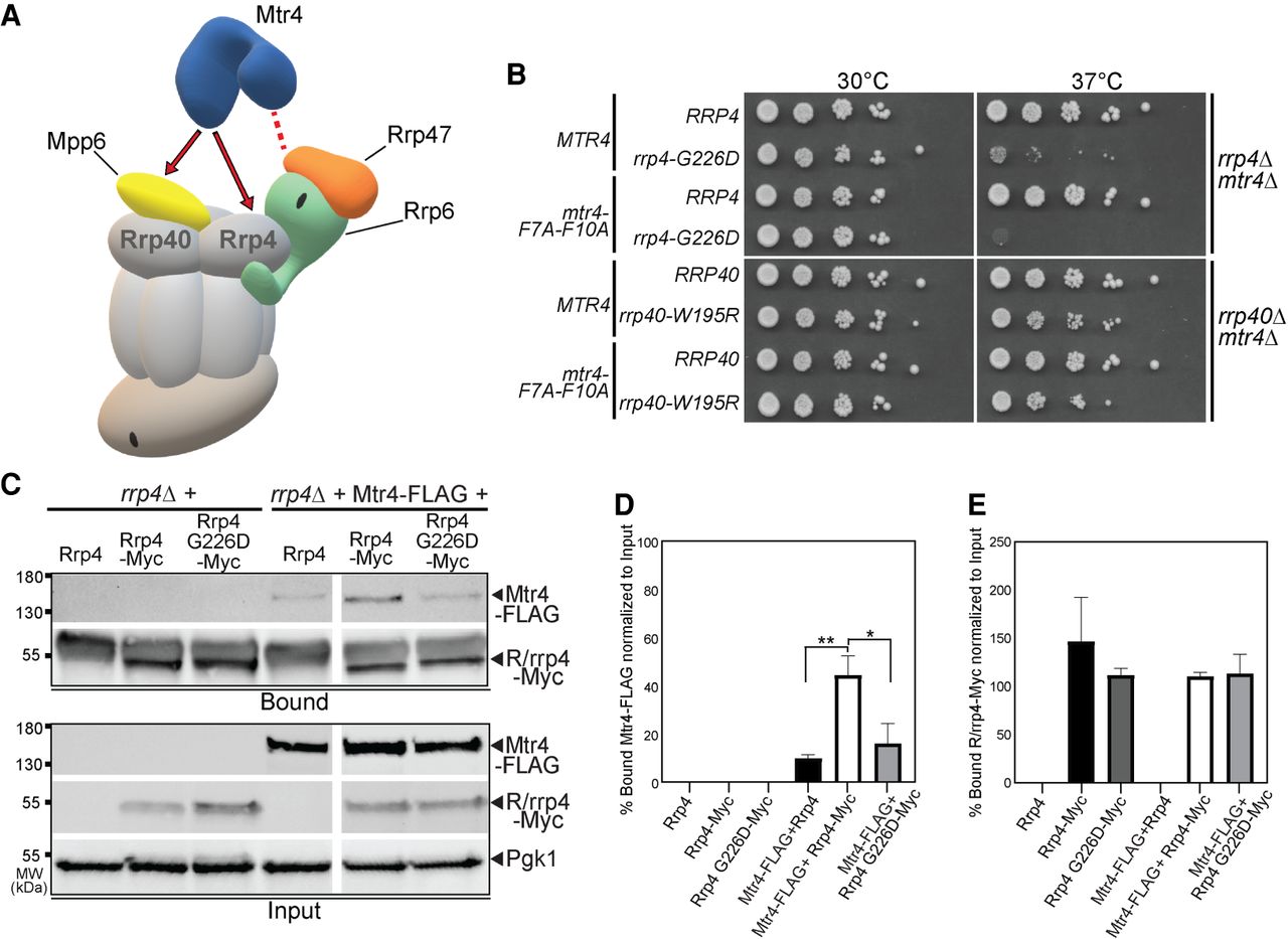
The rrp4-G226D mutant shows genetic interaction with an mtr4 mutant that is impaired for interaction with Rrp6/Rrp47 and Rrp4 G226D impairs interaction with Mtr4. (A) Cartoon of the budding yeast nuclear RNA exosome depicting the molecular interactions that the essential RNA helicase, Mtr4, makes with the RNA exosome, and exosome cofactors (Schuch et al. 2014; Falk et al. 2017; Schuller et al. 2018). The association of the amino terminus of Mtr4 with the RNA exosome is facilitated by interactions with nuclear exosome cofactors, Rrp6/Rrp47 (denoted by the dashed red line). The association of Mtr4 with the RNA exosome is also facilitated by interactions with nuclear exosome cofactor, Mpp6, which is associated with the Rrp40 exosome subunit, and the Rrp4 exosome subunit (denoted by the solid red arrows). (B) Double mutant cells containing rrp4-G226D and mtr4-F7A-F10A, an mtr4 mutant impaired for interaction with Rrp6/Rrp47, show lethality compared to the impaired growth of the single mutant rrp4-G226D at 37°C. In contrast, double mutant cells containing rrp40-W195R and mtr4-F7A-F10A show impaired growth at 37°C that is similar to the single mutant rrp40-W195R, which has been described previously (Fasken et al. 2017; Gillespie et al. 2017). The rrp4Δ mtr4Δ double mutant cells containing RRP4 or rrp4-G226D plasmid and rrp40Δ mtr4Δ double mutant cells containing RRP40 or rrp40-W195R plasmid that also harbor MTR4 or mtr4-F7A-F10A plasmid were serially diluted, spotted onto solid media, and grown at the indicated temperatures for 3 d. (C) The Rrp4 G226D variant shows decreased association with Mtr4. Myc-tagged Rrp4 and Rrp4 G226D protein was immunoprecipitated from rrp4Δ cells coexpressing Rrp4-Myc and FLAG-tagged Mtr4 grown at 30°C using anti-Myc beads, and the amount of bound Mtr4-FLAG protein was detected by immunoblotting. The bound/input level of Mtr4-FLAG was detected with an anti-FLAG antibody, and the bound/input level of Rrp4-Myc was detected with an anti-Myc antibody. The input level of 3-phosphoglycerate kinase (Pgk1) was detected as a loading control. (D) Quantitation of the percentage of bound Mtr4-FLAG coimmunoprecipitated with Rrp4-Myc and Rrp4 G226D-Myc. Graph shows the mean percentage of bound Mtr4-FLAG from three independent experiments (n = 3). Error bars represent standard error of the mean. Statistical significance is denoted by asterisks (*P-value ≤0.05; **P-value ≤0.01). (E) Quantitation of percentage of bound Rrp4-Myc and Rrp4 G226D-Myc immunoprecipitated. Error bars represent standard error of the mean. The coimmunoprecipitations were performed and quantitated as described in Materials and Methods.




