


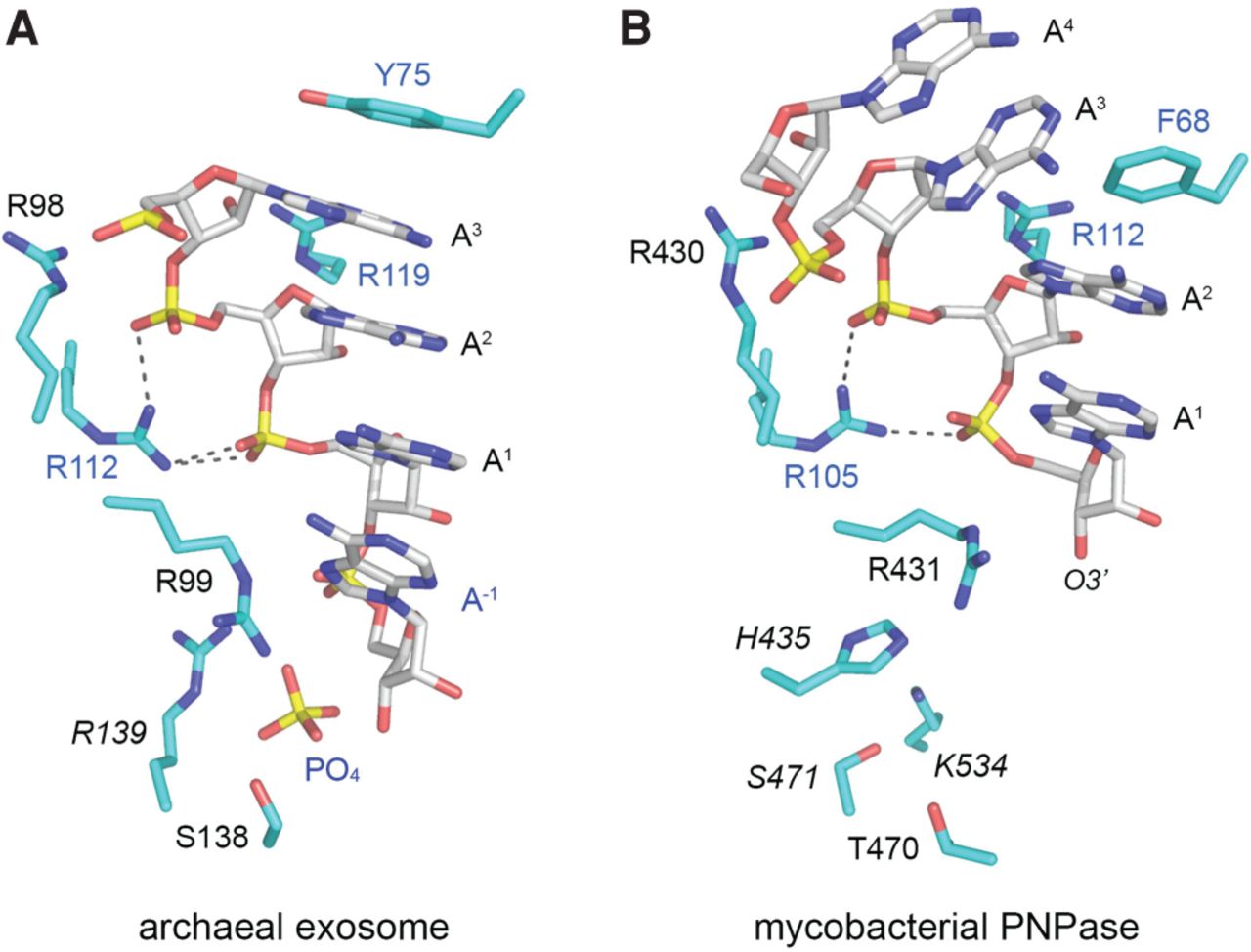
Comparison of RNA-bound mycobacterial and archaeal PNPase structures. The MsmPNPase•RNA complex (B) was superimposed on the structure of a Sulfolobus exosome(PNPase)•RNA•PO4 complex (A) and then offset laterally. The RNA ligands are shown as stick models with gray carbons and yellow phosphorus atoms. The adenylate nucleotides in the MsmPNPase complex are numbered from the 3′ end. The same numbering scheme is applied to the RNA in the Sulfolobus exosome structure, which has an additional 3′-terminal nucleotide (labeled A−1 in blue font). The phosphate anion in the archaeal exosome active site is shown as a stick model. Amino acids are depicted as stick models with cyan carbons. Amino acids from the Rrp42 subunit of the archaeal exosome and the proximal PH domain of MsmPNPase are labeled in blue font. Amino acids from the Rrp41 exosome subunit and the distal PH domain of MsmPNPase are labeled in black font. Contacts of the homologous Arg112/Arg105 side chains to adjacent phosphates of the RNA are indicated by dashed lines.




