


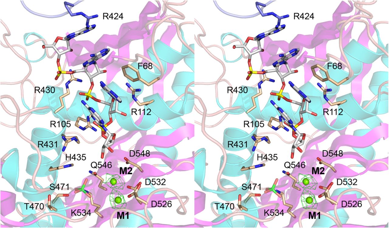
Stereo view of the MsmPNPase active site. PNPase protomer A is depicted as a cartoon model colored according to secondary structure (cyan α helices; magenta β strands). The (rA)4 tetranucleotide is rendered as a stick model with gray carbons and yellow phosphorus atoms. Magnesium ions M1 and M2 are depicted as green spheres within their density (green mesh, contoured at 2 σ, with 2 Å carve radius). A phosphate anion imported from the superimposed structure of E. coli PNPase is included as a semitransparent stick model with a green phosphorus atom, situated to the left of the metal ions. Amino acid side chains of protomer A that interact with the active site ligands are shown as stick models with beige carbons. The Arg424 side chain of a neighboring protomer that is situated adjacent to the 5′-terminal adenosine of (rA)4 is shown as a stick model with blue carbons.




