


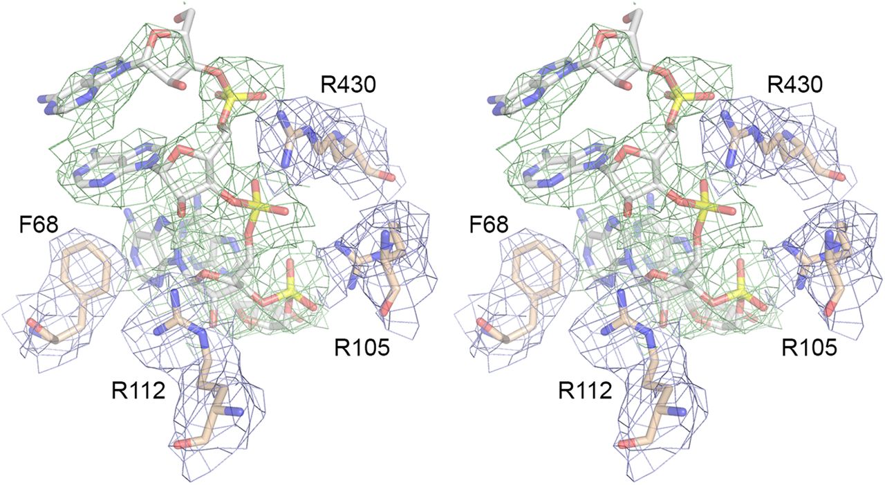
FIGURE 3.
Active site RNA ligand modeled into cryo-EM density. Stereo view of the (rA)4 tetranucleotide (stick model with gray carbons and yellow phosphorus atoms) modeled into surrounding density (green mesh, contoured at 1.25 σ, with 2 Å carve radius). Adjacent amino acids Phe68, Arg105, Arg112, and Arg430 are depicted as stick models with beige carbons modeled into their density (blue mesh, contoured at 2 σ, with 2 Å carve radius).




