


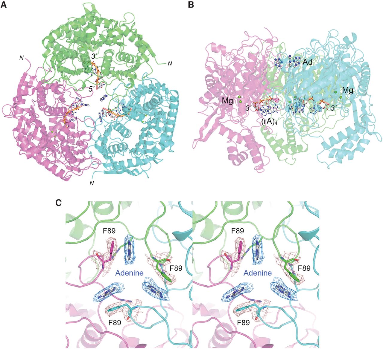
MsmPNPase in the act of poly(A) synthesis. The MsmPNPase homotrimer is depicted as a cartoon model, colored by protomer and oriented so that the top surface of the trimer is facing forward (A) or rotated to provide a side view (B). The r(A)4 RNA in the active site of each protomer is shown as a stick model with a gold cartoon trace through the phosphate backbone. The 5′ and 3′ ends of the RNA are labeled in panel A. The 5′ end is pointed toward the central channel; the 3′ end is directed toward the active site. Two magnesium ions in each active site are depicted as green spheres and labeled in panel B. The adenine nucleobase (Ad) in the central channel is rendered as a ball-and-stick model. (C) Close-up stereo view of the central channel looking down from the top of the PNPase trimer, highlighting how the adenine nucleobase (stick model with gray carbons) makes a π-stack on Phe89 (stick model, colored by protomer). The cryo-EM densities for adenine (blue mesh) and Phe89 (beige mesh) are shown (contoured at 2 σ, with 2 Å carve radius). Arg95 projecting into the central channel below Phe89 is also shown as a stick model.




