


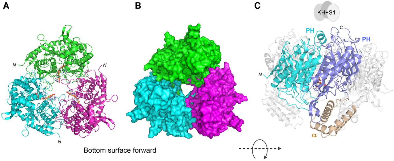
CryoEM structure of MsmPNPase. The MsmPNPase homotrimer is depicted as a cartoon model (A) and a surface model (B), colored by protomer and oriented so that the inferior/bottom surface of the trimer is facing forward. The r(A)4 RNA is shown in panel A as a stick model with a gold cartoon trace through the phosphate backbone. The structure is rotated in panel C to feature the component domain modules of the core PNPase protomer, wherein the PH domains are colored cyan and blue, and the intervening α-helical module is colored beige. The KH-S1 modules that could not be modeled into density are shown as a trio of gray ovals hovering above the carboxyl termini of the PNPase core structure.




