


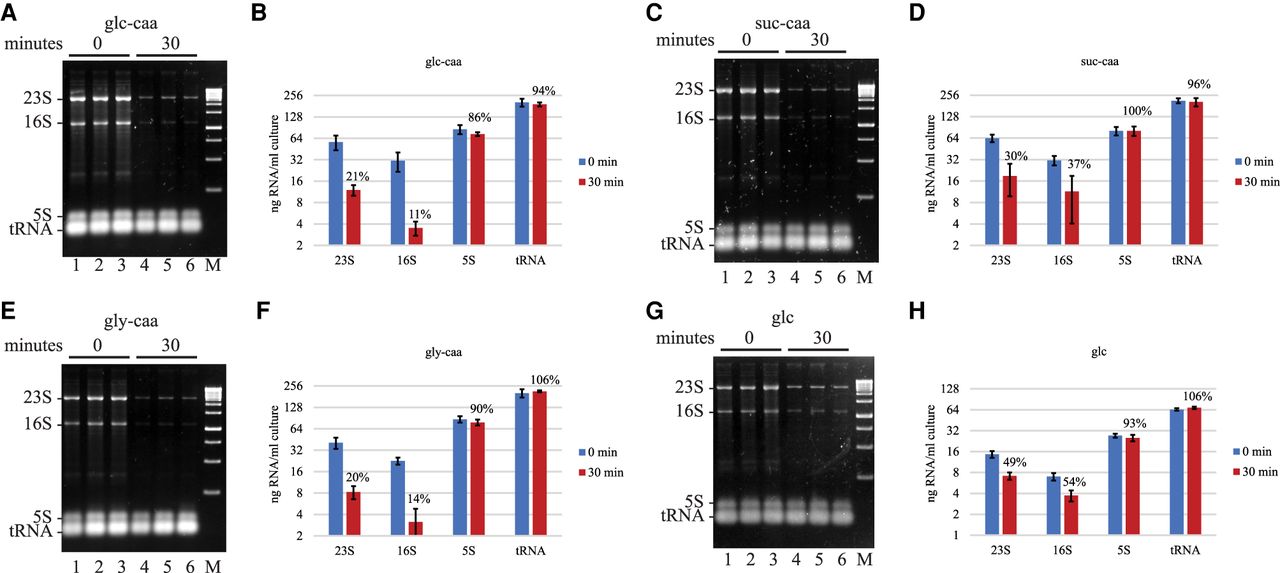
Rifampicin-induced rRNA degradation in MOPS media supplements with carbon sources and casamino acids. (A,C,E,G) SYBR Safe stained agarose gels. E. coli strain NCM3416 was grown in MOPS medium at 37°C supplemented with glucose (glc), succinate (suc) or glycerol (gly), and casamino acids (caa), except in G. Final concentration of carbon source and casamino acids was 0.5% and 0.2%, respectively. Each gel shows three biological replicates. The 0 min time point was taken immediately after the addition of rifampicin. (B,D,F,H) Levels of 23S, 16S and 5S rRNA, and tRNA were quantified from six biological replicates. Nominal amounts of RNA were determined as described (Fig. 2). The graphs show the average and standard deviation expressed as nanogram of RNA per mL of culture. Percentages represent normalization of the 30 min time point after rifampicin addition to the 0 min time point.




