


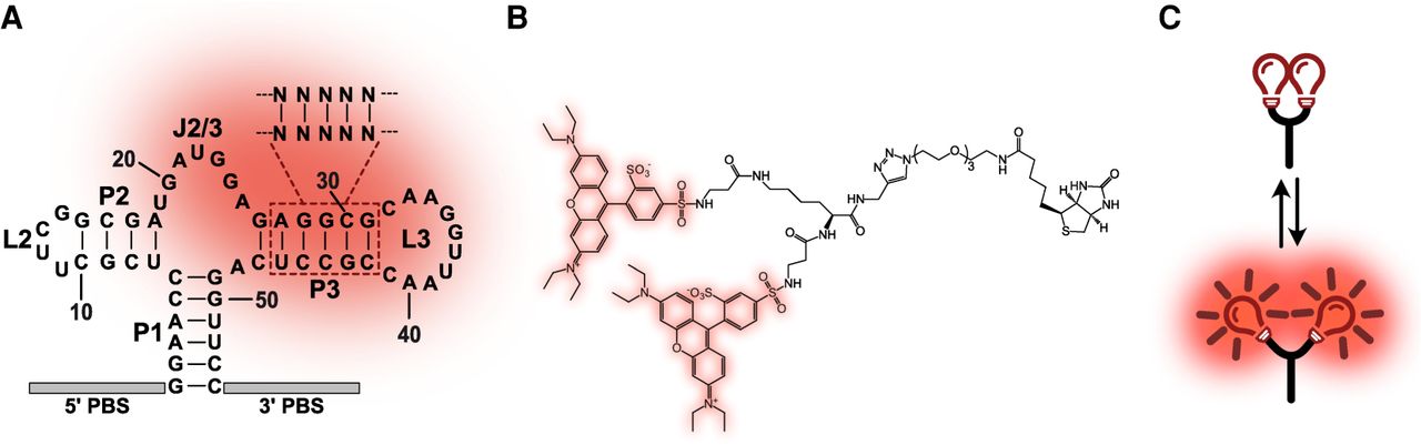
Structure and mode of action of SRB-2 RNA aptamer and Gemini-561 fluorogen. (A) SRB-2 aptamer and P3 mutant library. The molecule is shown as initially described (Holeman et al. 1998). The 10 positions randomized in the P3 stem are dashed-boxed and the domains proposed to interact with sulforhodamine B are shadowed in red. SRB-2 and its derivatives were flanked with two constant regions used as primer binding sequences (PBS) for PCR amplifications. (B) Gemini-561 fluorogen. The fluorogen is made of a dimer of sulforhodamine B (shadowed in red) linked together by a lysine and a PEG linker. The fluorogen also carries a biotin moiety initially used for aptamer selection (Bouhedda et al. 2020). (C) Working principle of Gemini-561. H-aggregates form when the molecule is dissolved in an aqueous environment, leading to sulforhodamine B fluorescence quenching. However, modulating medium polarity (e.g., by adding methanol) or in the presence of SRB-2 aptamers, both sulforhodamine B moieties separate and recover their fluorescence capacity.




