


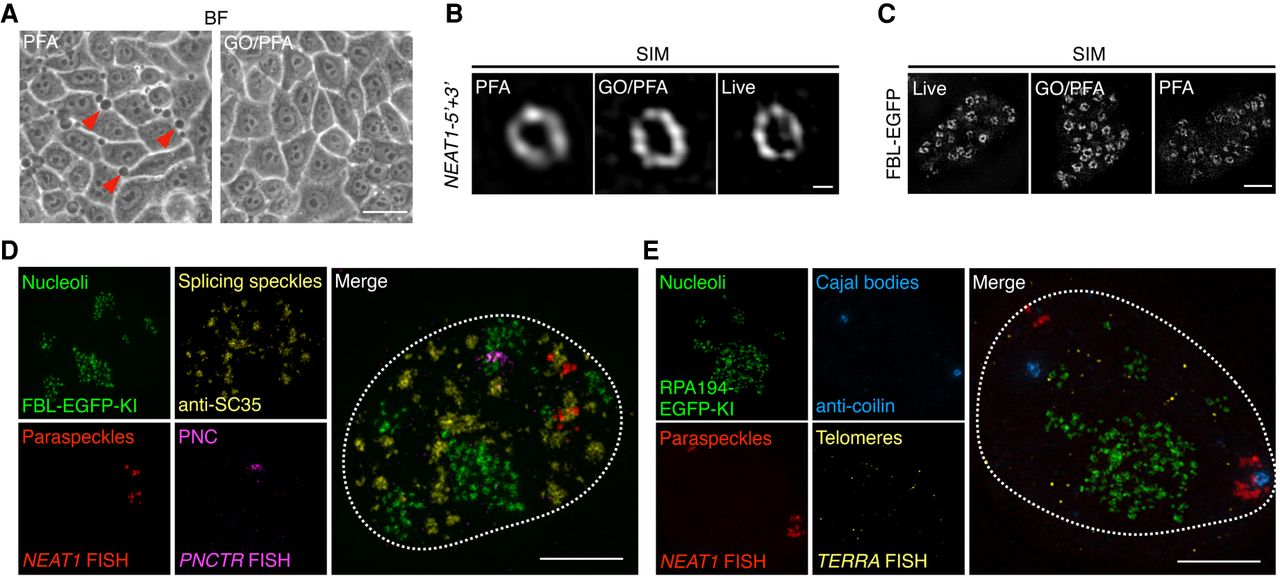
Glyoxal/PFA fixation enables the covisualization of different types of nuclear bodies with minimal impact on their ultrastructures. (A) GO/PFA fixation shows a better preservation of the cell morphology than PFA alone, shown by fewer membrane blebbing (red arrows, left). BF, bright field. Scale bar: 15 µm. (B) Structured illumination microscopy (SIM) reveals that GO/PFA fixation enables a better preservation of the shell of paraspeckles labeled by NEAT1-5′ + 3′ RNA FISH than PFA alone. Scale bar: 200 nm. (C) SIM reveals that GO/PFA fixation enables a better preservation of the ultrastructure (the DFC regions) of nucleoli labeled by the CRISPR/Cas9-mediated EGFP knock-in at the endogenous FBL locus. Scale bar: 2 µm. (D,E) GO/PFA fixation enables the covisualization of different types of nuclear bodies. Nucleoli were labeled by the CRISPR/Cas9-mediated EGFP knock-in at the endogenous FBL locus (D) or CRISPR/Cas9-mediated EGFP knock-in at the endogenous RPA194 locus (E); paraspeckles were labeled by lncRNA NEAT1 FISH; perinucleolar compartments (PNC) were labeled by the lncRNA PNCTR FISH; telomeres were labeled by lncRNA TERRA FISH; splicing speckles were labeled by anti-SC35 antibodies; Cajal bodies were labeled by anticoilin antibodies. Nuclei were marked by dotted line. Scale bar: 5 µm.




