


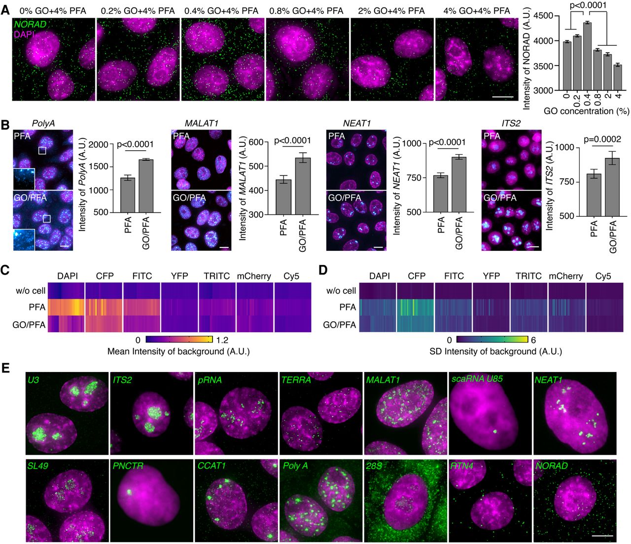
Addition of glyoxal improves RNA FISH signals in nuclear bodies. (A) Introducing 0.4% glyoxal into 4% PFA improves the RNA FISH signals. Statistics were derived from over 4000 NORAD molecules counted across three independent experiments under each condition. Mean ± SEM and Mann–Whitney test are shown. Scale bar: 10 µm. (B) GO/PFA fixation dramatically enhances the FISH signals for different kinds of RNAs (cyan) in nuclei (magenta). Note that cytoplasmic polyadenylated RNAs could be readily detected by GO/PFA fixation; but these RNAs were barely detected by the PFA alone fixation (first panel, enlarged view). Statistics were counted from more than 100 cells under each condition. Mean ± SD and Mann–Whitney test are shown across three independent experiments for each RNA. Scale bar: 5 µm. (C) Glyoxal does not cause additional autofluorescence in all commonly used channels. The heat map was generated from 30 images for each channel under each fix condition. (D) Glyoxal provides a uniform background in all commonly used channels. The heat map was generated from 30 images for each channel in each fix condition. (E) GO/PFA fixation provides high-quality FISH images for different types of RNAs. Nuclei were labeled by DAPI and shown in magenta. Scale bar: 5 µm.




