


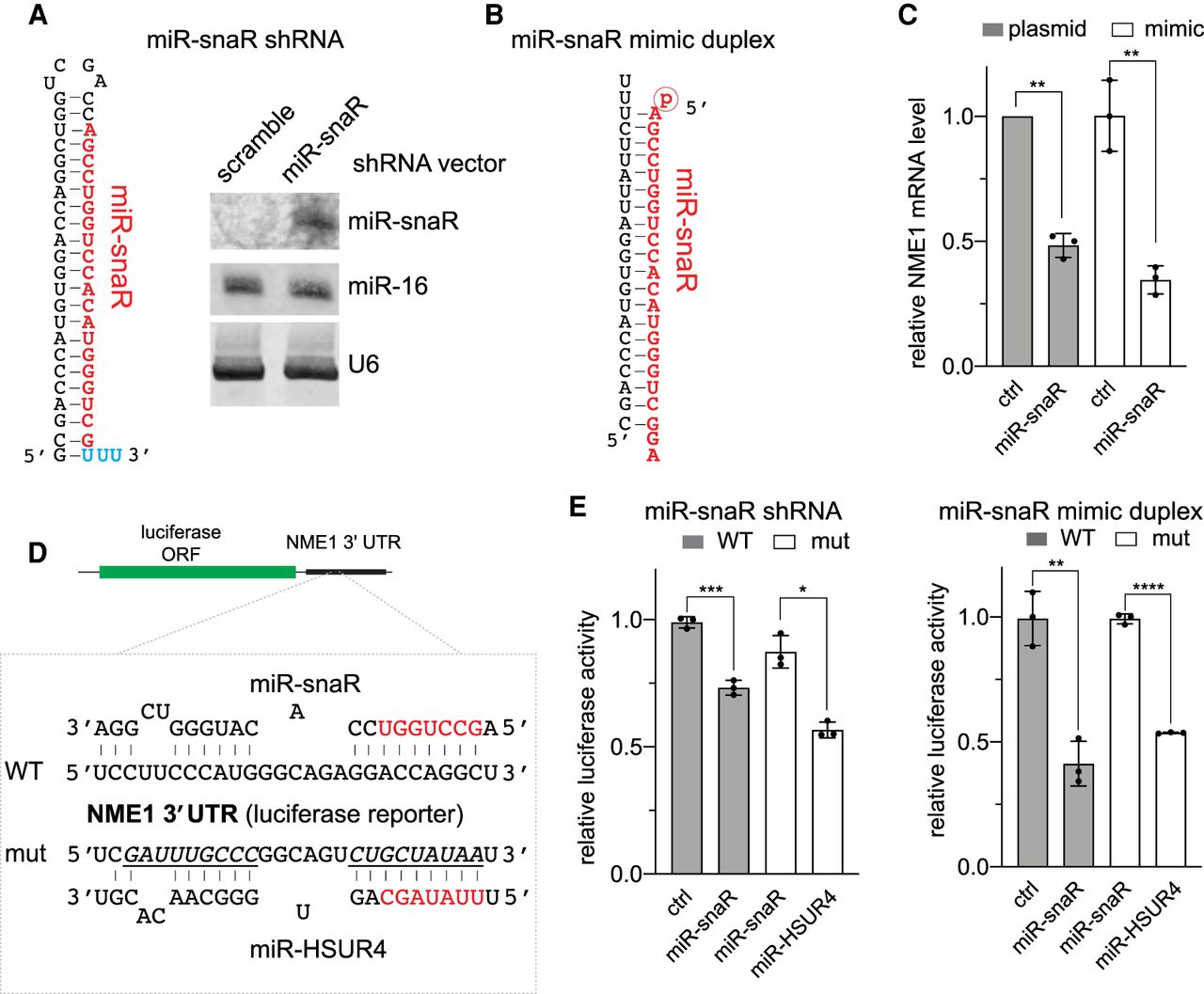
miR-snaR represses NME1 mRNA by interacting with its 3′-UTR. (A, left) Schematic of the shRNA encoded in pU6-miR-snaR, with miR-snaR sequence highlighted in red. The terminal Us (cyan) are required for efficient RNA Pol III termination. (Right) Northern blot analyzing the levels of miR-snaR in total RNA extracted from 293T cells transfected with either pU6-scramble-shRNA or pU6-miR-snaR. Endogenous miR-16 and U6 serve as internal loading controls. (B) Schematic of the miR-snaR mimic duplex. The miR-snaR mimic strand in red is modified with a 5′ phosphate group. (C) RT-qRCR analysis of NME1 mRNA in total RNA extracted from 293T cells transfected with pU6-miR-snaR or miR-snaR mimic. NME1 mRNA expression was normalized to β-actin. Three biological replicates for each sample were recorded and data was graphed and analyzed using Prism GraphPad. (**) P ≤ 0.01. (D) Depiction of WT and mutant NME1 target sites on the luciferase reporter along with their targeting miRNA: miR-snaR and miR-HSUR4, respectively. Seed sequences of the miRNAs are highlighted in red. Regions mutated in the mutant reporter are underlined. (E) Dual luciferase reporter containing the WT or mut NME1 3′-UTR downstream from firefly luciferase was transfected in 293T cells together with either pU6-miR-snaR (left) or miR-snaR mimic (right). Firefly luciferase activity was measured 48 h post transfection and normalized to Renilla luciferase activity. Error bars represent standard deviation from three experiments. All differences are statistically significant (Student's t-test, [*] P ≤ 0.05; [**] P ≤ 0.01; [***] P ≤ 0.001; [****] P ≤ 0.0001).




