


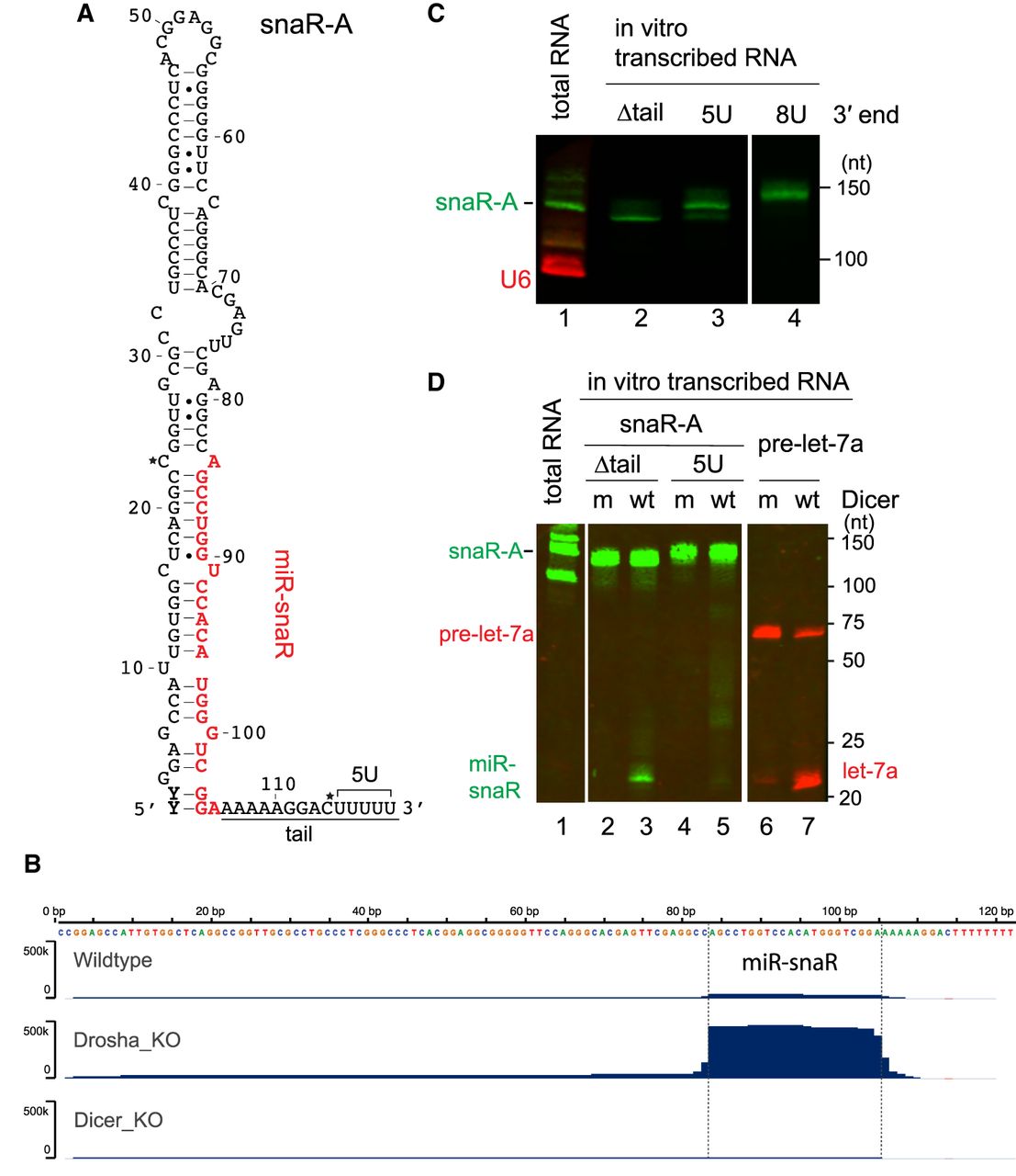
miR-snaR can be released from a truncated snaR-A transcript by Dicer. (A) Predicted secondary structure of snaR-A1, with the miR-snaR sequence highlighted in red. The two nucleotides that are variable within the 14 snaR-A family members are marked by a star. (B) Histogram showing small RNA reads mapped to the snaR-A1 locus. Reads are identified in Ago-qCLASH experiments from HCT116 WT, Drosha-KO, and Dicer-KO cells. The boundaries of the miR-snaR are indicated by dotted lines. (C) Northern blot detection of various in vitro transcribed snaR-A transcripts, as well as endogenous snaR-A and U6 in total RNA extracted from 293T cells (nt: nucleotides). (D) In vitro Dicer processing assay. In vitro transcribed pre-let-7a and snaR-A with or without the tail (illustrated in A) were processed by purified flag-tagged Dicer and analyzed by northern blot (wt: wild-type Dicer.; m: Dicer with mutated RNase III domains).




