


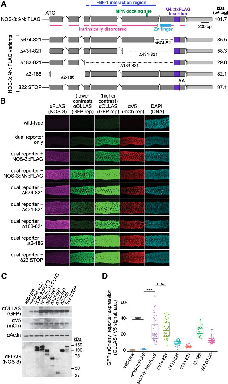
Analysis of tethered NOS-3 deletion mutants for enhancement of reporter expression. (A) nos-3 locus and variants used to identify the region necessary for reporter expression enhancement. Variants include four in-frame deletions and a premature STOP codon that removes amino acids carboxy-terminal to the tag insert. Variant protein sizes (kilodaltons, kDa) shown at right. Coding regions, gray; untranslated regions, light gray; λN::FLAG insert, purple. Intrinsically disordered regions were predicted by IUPred2A (Erdos and Dosztanyi 2020). FBF-1 interacting region (Kraemer et al. 1999) and MPK docking site (Arur et al. 2011) labeled as previously identified. (B) Confocal images (max projection) of adult germlines. Conventions as detailed in Figure 1D. See Figure 1B for germline image location. Note that images for wild-type, reporter only, NOS-3::FLAG, and NOS-3:: λN::FLAG are replicated from Figure 1D. (C) Immunoblot to assay expression of reporter proteins (above) and NOS-3 variant proteins (below). Conventions as in Figure 1F. SDS-PAGE sizes (kDa) shown to right for the NOS-3 immunoblot. Whole worm lysate (n = 60 young adult worms) used for all samples. (D) Relative abundance of GFP and mCherry reporter proteins. Ratio of signals from GFP (αOLLAS) and mCherry (αV5) reporter proteins, averaged across distal 100 µm of germline and normalized to wild-type negative control. Wild-type (N2, no dual reporter; n = 54), NOS-3::FLAG (n = 34), NOS-3:: λN::FLAG (n = 52), Δ183–821 (n = 37), Δ431–821 (n = 36), Δ674–821 (n = 42), Δ2–186 (n = 31), 822 STOP (n = 37) worms analyzed for B and D. Curves and values of wild-type, NOS-3::FLAG and NOS-3:: λN::FLAG are same as reported in Figure 1E and Supplemental Figure S2. (***) P-value <0.0001. n.s., not significant (P-value = 0.5735). All P-values for comparisons between strains can be found in Supplemental Table S1.




