


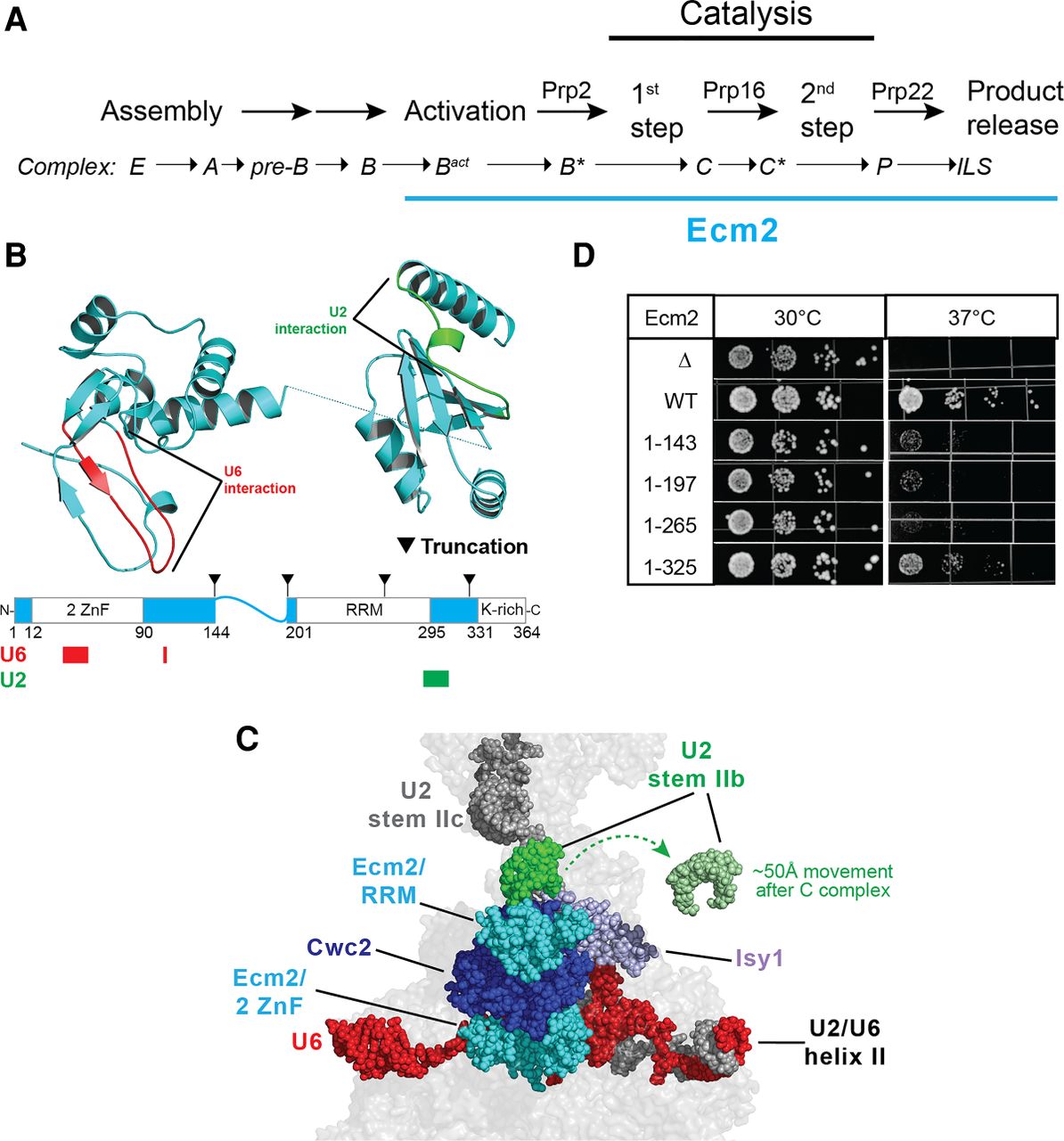
Structural analysis of Ecm2 during splicing. (A) Schematic of the pre-mRNA splicing pathway. ATPases tested for genetic interactions with Ecm2 are shown above the arrows of the respective steps that they promote. Spliceosome complexes containing Ecm2 are underlined in blue. (B) Cryo-EM structure and domain organization of Ecm2. U6 and U2 snRNA interacting regions are marked and shown in red and green, respectively. Locations of truncation mutants studied in panel C are indicated. Structure from 6EXN.pdb. (C) Structure of the Cwc2/Ecm2/Isy1 hub and Ecm2-RRM/U2 stem II interaction in C complex. The position of stem IIb after remodeling in C* complex has been superimposed on this structure. The U6 snRNA, Cwc2, and Ecm2 do not significantly change positions in C* complex. This figure was created using 5LJ5.pdb and 5MQ0.pdb. (D) Temperature sensitivity of ecm2Δ and truncation mutants on -ura dropout media after 3 days of growth.




