


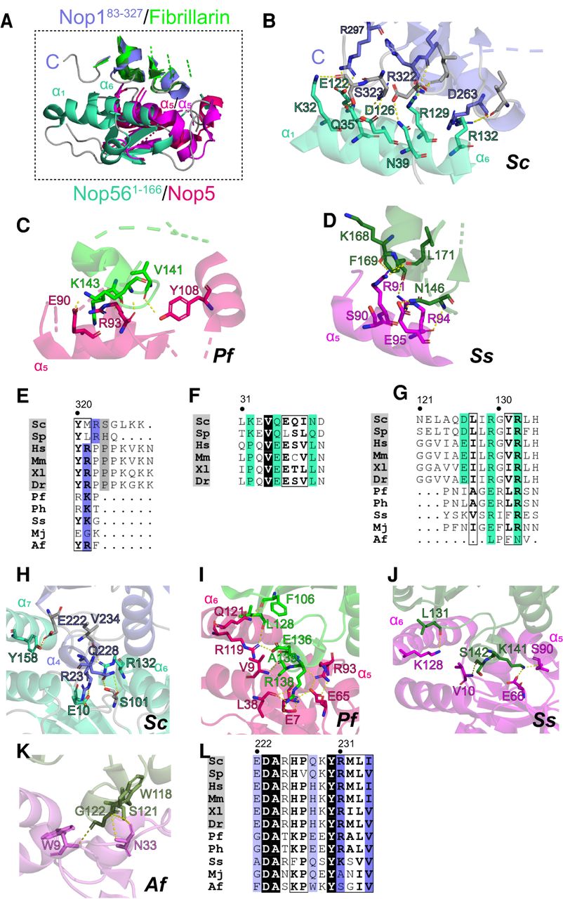
Comparison of interaction hot-spots in the eukaryotic Nop561–166–Nop183–327 complex and ortholog archaeal complexes. (A) Expansion of the region comprising Nop56-α1 and the carboxy-terminal tail of Nop1 of the Sc Nop561–166–Nop183–327 complex overlayed with archaeal orthologous Nop5–Fib complexes. Sc Nop183–327, Pf and Ss Fibrillarin are in slate, green and forest, respectively; Sc Nop561–166, Pf and Ss Nop5 are in green cyan, hot pink, and magenta, respectively. The Pf and Ss structures are from PDB-IDs 2NNW and 3ID5, respectively. (B) Contacts between Nop561–166-α1 and -α6 and Nop1. (C) Contacts between Pf Nop5-α5 and Pf Fibrillarin. (D) Contacts between Ss Nop5-α5 and Ss Fibrillarin. Hydrogen bonds are depicted as yellow dashed lines. (E) Multiple sequence alignment of the carboxy-terminal residues of eukaryotic and archaeal Fibrillarin; conserved residues are highlighted in slate. (F) Multiple sequence alignment of Nop56-α1 in eukaryotes. Interacting residues in S. cerevisiae are highlighted in green cyan. (G) Multiple sequence alignment of eukaryotic Nop56-α6 and archaeal Nop5-α5. Interacting residues are highlighted in green cyan. (H) Contacts between Nop183–327-α4 and Nop561–166. (I) Contacts between Pf Fib-α4 and Pf Nop5. (J) Contacts between Ss Fib-α4 and Ss Nop5 (K) Contacts between Af Fib-α4 and Af Nop5. (L) Multiple sequence alignment of α4 residues of eukaryotic and archaeal Fibrillarins; conserved and interacting residues are highlighted in slate. Residue numbers refer to the first line of the alignment.




